by Rosemarie Tremblay-LeMay MD MSc FRCPC and Vathany Kulasingam, PhD, FCACB
March 2, 2022
What is a plasma cell neoplasm?
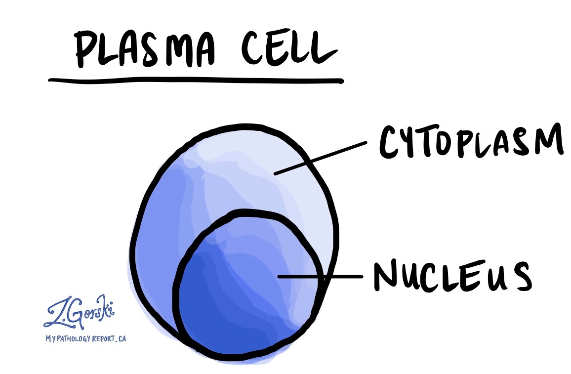
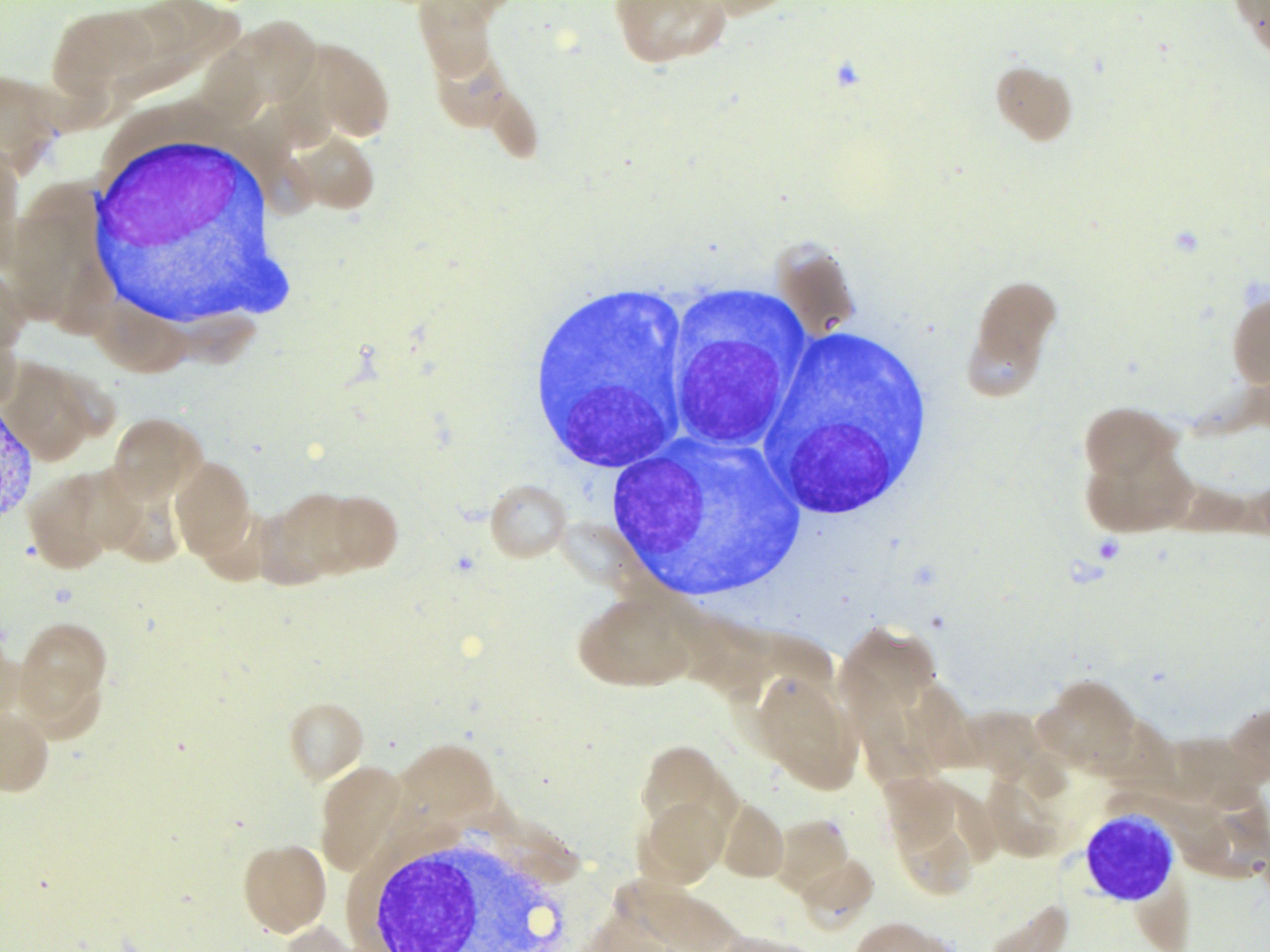
A plasma cell neoplasm is a type of cancer that starts from specialized immune cells called plasma cells. Normal plasma cells make different types of proteins called immunoglobulins (antibodies) that help protect the body from micro-organisms such as viruses. In contrast, all of the plasma cells in a plasma cell neoplasm make the same type of immunoglobulin. The abnormal plasma cells also make a lot more immunoglobulin than normal plasma cells.
What are plasma cells?
Plasma cells start off life as a specialized type of immune cell called B-lymphocytes. Once a B-lymphocyte turns into a plasma cell, it has the ability to produce special proteins called immunoglobulins (Ig), which are also called antibodies. Immunoglobulins protect our body by sticking to bacteria and viruses, which makes them easier to remove from the body. Immunoglobulins can also stick to abnormal cells or cells that have stopped functioning normally.

Immunoglobulins are made up of four parts and each part is called a chain. One immunoglobulin is made up of two heavy chains and two light chains. There are five different kinds of heavy chains, called A, G, D, E, M, and two different kinds of light chains called kappa and lambda. Any combination of heavy and light chains can be used to make an immunoglobulin. These options allow your body to produce many different kinds of immunoglobulins (for example IgA kappa, IgG lambda, etc.).
While the immune system has the ability to make many different kinds of immunoglobulins, each plasma cell makes just one kind of immunoglobulin. Because our immune system makes millions of different plasma cells, it is normal to find many different kinds of immunoglobulins in the body at any time.
What tests do doctors perform to look for abnormal immunoglobulins?
Your doctor can order a test called serum protein electrophoresis to see the immunoglobulins in your blood or urine. The results of the test appear as a line on a graph. A normal result looks like a line with many small bumps. Each small bump in the line is a different type of immunoglobulin. An abnormal result shows a large peak (see the black area in the image below). This large peak is the abnormal immunoglobulin. Other tests can show the accumulation of light chains in your blood or urine.
The abnormal plasma cells in a plasma cell neoplasm will produce only one type of immunoglobulin. This will result in increased amounts of either IgG or IgA (or rarely IgD or IgE) and an excessive amount of either kappa or lambda compared to normal.

How do pathologists make the diagnosis of plasma cell neoplasm?
The diagnosis of plasma cell neoplasm is usually made after your doctor takes a small piece of your bone marrow in a procedure called a biopsy. For some patients, the abnormal plasma cells form a tumour outside of the bone. In that situation, your doctor may perform a biopsy of that tumour instead. Rarely, the plasma cells can be seen in your blood. The tissue is then sent to your pathologist who examines it under the microscope.
By examining the tissue under the microscope, your pathologist will determine the percentage of abnormal plasma cells present in your bone marrow. Your doctor will be able to combine this information with other test results to determine the type of plasma cell neoplasm.

What other tests do pathologists perform for plasma cell neoplasm?
Immunohistochemistry
Your pathologist will perform a test called immunohistochemistry to learn more about the plasma cells in your tissue sample and confirm that they are abnormal. Immunohistochemistry is a test that uses antibodies to highlight different types of proteins produced by cells. When the cells produce a protein, pathologists describe the result as positive or reactive. When the cells do not produce the protein, the result is described as negative or non-reactive.
The cancer cells in plasma cell neoplasms come from plasma cells and as a result, they produce proteins normally made by plasma cells, such as CD138, MUM1, or CD79a. They can also produce proteins that are not produced by normal plasma cells, such as CD20, CD117, CD56, or CyclinD1.
In situ hybridization
Your pathologist may also perform a test called in situ hybridization (ISH) to determine which immunoglobulins are being produced by the abnormal plasma cells. As described above, these abnormal plasma cells in a plasma cell neoplasm will produce only one type of immunoglobulin, for example, IgG kappa or IgG lambda.
Molecular tests
Each cell in your body contains a set of instructions that tell the cell how to behave. These instructions are written in a language called DNA and the instructions are stored on 46 chromosomes in each cell. Because the instructions are very long, they are broken up into sections called genes and each gene tells the cell how to produce a piece of the machine called a protein.
Sometimes, a piece of DNA falls off one chromosome and becomes attached to a different chromosome. This is called translocation and it can result in the cell making a new and abnormal protein. If the new protein allows the cell to live longer than other cells or spread to other parts of the body, the cell can become cancer. The malignant cells can also lose or gain a piece of DNA.
Pathologists usually test for these molecular changes by performing fluorescence in situ hybridization (FISH) on a piece of the tissue from the tumour. This type of testing can be done on the biopsy specimen.
Different types of abnormalities can be seen in plasma cell neoplasms. They include translocations involving the protein IGH, loss of chromosome 1p or 17p (codes for the gene TP53), and gain of chromosome 1q. The presence of these abnormalities can help your doctor determine your prognosis.
How do the abnormal plasma cells in a plasma cell neoplasm damage the body?
There are three ways that a plasma cell neoplasm damages the body:
- Kidney injury – The kidneys are responsible for removing waste and harmful chemicals from your blood. The kidneys normally remove immunoglobulins from the blood, but high levels of immunoglobulins can damage your kidneys.
- Bone damage – The abnormal plasma cells can build up inside bones. This results in bone damage and an increased amount of calcium in the blood.
- Decreased red blood cells (anemia) – The production of red blood cells depends on both healthy kidneys and bone marrow. As a result, patients with a plasma cell neoplasm often produce fewer red blood cells because of the damage to their kidneys and bones. This condition is called anemia.
What are the types of plasma cell neoplasms?
Plasma cell neoplasm can be divided into different categories based on how much immunoglobulin is found in your blood or urine, as well as how many plasma cells are seen in your bone marrow and whether or not there is evidence of damage to your organs.
- Monoclonal gammopathy of undetermined significance (MGUS) – This diagnosis is made when plasma cells represent less than 10% of the cells in the bone marrow, the immunoglobulins detected in the blood or urine are below a certain threshold, and there is no damage to your organs. MGUS is considered a pre-cancerous condition because only a small percentage of persons with this condition will progress to the next stage.
- Plasma cell myeloma – This diagnosis is made when there are more than 10% plasma cells in your bone marrow and the immunoglobulins found in your blood or urine are above a certain threshold.
- If there is no evidence of damage to your organs, this is called an asymptomatic (or smoldering) plasma cell myeloma.
- If there is evidence of damage to your organs or more than 60% of plasma cells in your bone marrow, this is called a plasma cell myeloma or multiple myeloma.
What is a plasmacytoma?
Sometimes the abnormal plasma cells can group together to form a tumour. A tumour made up of abnormal plasma cells is called a plasmacytoma. When a plasmacytoma forms outside the bone it is called an extraosseous plasmacytoma. If only a single tumour is found in a bone without injury to other parts of the body, it is called a solitary plasmacytoma of the bone.
What is plasma cell leukemia?
The abnormal plasma cells can also go into the blood. If they represent more than 20% of the white blood cells in the blood, it will be called plasma cell leukemia.
What is amyloidosis?
Sometimes, the abnormal immunoglobulins produced by the plasma cells will build up in tissues. When this happens, it can create a substance called amyloid. Amyloidosis is a condition where large amounts of amyloid build up in the body and cause damage to organs. In amyloidosis, there may only be a small population of abnormal plasma cells seen in the bone marrow. In order to see amyloid, your pathologist can use a special stain called Congo Red. Using this stain the amyloid looks red under normal light and apple green under a special light.

