

Keratinizing dysplasia is a precancerous condition marked by an abnormal growth pattern in squamous cells. These cells are found on external and internal body surfaces such as the skin, mouth, throat, and cervix.
Dysplasia means the cells look abnormal under the microscope and are not maturing or developing normally. In keratinizing dysplasia, these abnormal squamous cells also produce excessive amounts of keratin, a protective protein normally found on the skin’s outer layer.
Is keratinizing dysplasia a precancerous change?
Yes, keratinizing dysplasia is considered a precancerous condition. This means the abnormal cells have the potential to develop into squamous cell carcinoma, a type of cancer. However, not all cases of keratinizing dysplasia will progress to cancer. Regular medical follow-ups and treatment can help manage this risk.
What causes keratinizing dysplasia?
The cause of keratinizing dysplasia varies depending on the location in the body. Common causes include smoking, heavy alcohol use, or long-term inflammation.
What areas of the body are affected by keratinizing dysplasia?
Keratinizing dysplasia typically occurs in areas with squamous cells that line moist surfaces of the body, known as mucosal surfaces. Common locations include the mouth, tongue, throat (pharynx and larynx), esophagus, cervix, and genital area. It can also occasionally affect the skin, especially in regions experiencing chronic irritation.
Are there different grades of keratinizing dysplasia?
Yes, keratinizing dysplasia is graded based on how abnormal the cells appear under the microscope. The grades include:
- Mild dysplasia: Slight abnormalities in cells, low risk of becoming cancer.
- Moderate dysplasia: More noticeable cell abnormalities, moderate risk of progressing to cancer.
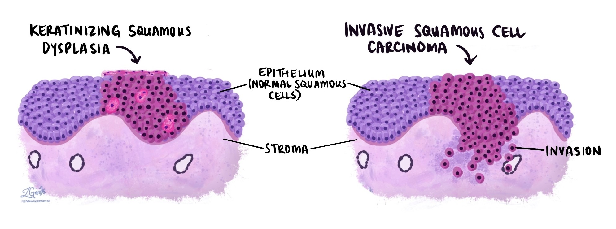
- Severe dysplasia: Marked cell abnormalities, high risk of progressing to squamous cell carcinoma. Severe dysplasia is sometimes called carcinoma in situ, meaning it has not yet invaded deeper tissues.
The grade helps doctors determine appropriate treatment and follow-up.
What does keratinizing dysplasia look like under the microscope?
Under a microscope, keratinizing dysplasia appears as abnormal squamous cells that are disorganized and produce large amounts of keratin. These cells often stain bright pink or red due to the keratin content. The abnormal cells may be found in thickened layers, and pathologists look for features such as increased cell size, irregular cell shapes, enlarged nuclei, and clumped keratin, known as keratin pearls. Recognizing these changes helps pathologists make the correct diagnosis.
Questions to ask your doctor
-
What grade of keratinizing dysplasia was found in my sample?
-
Does this mean I am at high risk of developing cancer?
-
What treatments or follow-up do you recommend for me?
-
Can lifestyle changes, such as quitting smoking or reducing alcohol, lower my risk?
-
How often should I have follow-up tests or examinations?
We are proud to partner with:
![]()