November 14, 2023

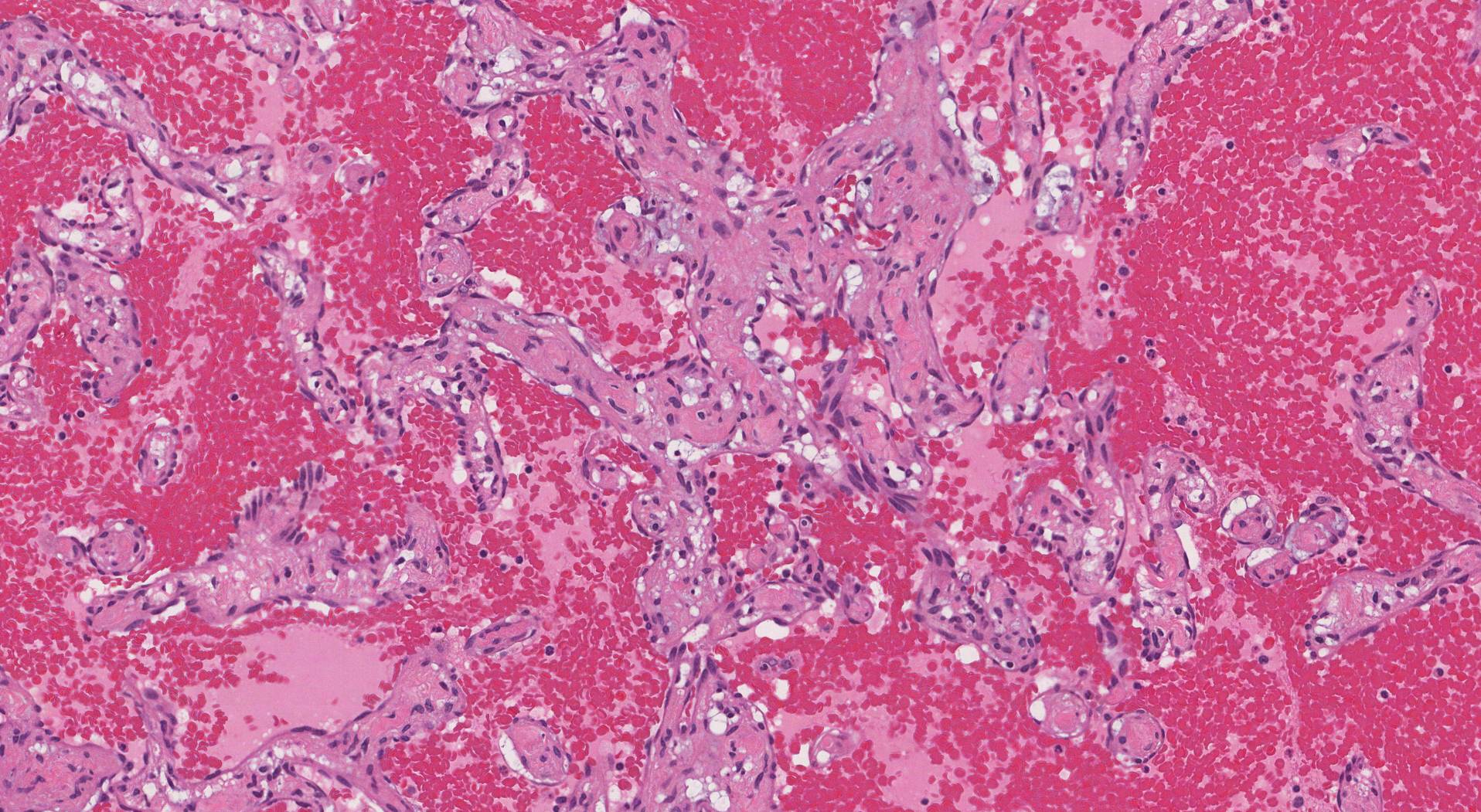
A vascular lesion is a growth predominantly composed of endothelial cells forming abnormal blood vessels. The term vascular lesion can be applied to a wide range of conditions, including benign (noncancerous) tumours, intermediate (locally aggressive) tumours, malignant (cancerous) tumours, congenital/development abnormalities, and reactive conditions. Vascular lesions can occur in any part of the body, although they are most commonly encountered in the skin.
Types of vascular lesions
Doctors often divide vascular lesions into one of five groups: reactive, congenital/developmental, benign (noncancerous), intermediate (locally aggressive), and malignant (cancerous).
Reactive vascular lesions
These lesions are called reactive because they respond to changes in the local tissue environment, such as an infection or injury. They are common lesions that can be found almost anywhere in the body. They are noncancerous growths.
- Granulation tissue: This is a type of tissue that develops after any injury. It often contains many small blood vessels that bring immune cells and nutrients to the injured area of the body. Although it is described as a lesion, it is part of the normal healing process.
- Lobular capillary hemangioma (pyogenic granuloma): This is a red, fleshy, and often bleeding bump that usually occurs on the lips, fingers, or toes. It may develop after a minor injury, during pregnancy, or while taking certain medications.
- Reactive angioendotheliomatosis: This is a rare condition that causes red or purple patches or nodules on the skin. It is associated with blood clots, infections, autoimmune diseases, or cancers.
- Stasis dermatitis: This condition causes purple or brown spots on the lower legs or feet. Chronic venous insufficiency, arteriovenous malformations, or trauma cause it.
- Bacillary angiomatosis: This condition is associated with red or purple papules or nodules on the skin or mucous membranes. It is caused by a bacterial infection, usually in immunocompromised patients.
Congenital/developmental vascular lesions
These lesions typically develop before or shortly after birth, although they may not become noticeable until later in life. They are noncancerous growths.
- Capillary malformations: These lesions are typically found in the skin where they are also known as port-wine stains or salmon patches. They are flat red or pink patches of skin that are caused by dilated capillaries. They are usually present at birth and may grow larger or darker over time. They can occur anywhere on the body but are most common on the face, neck, and limbs.
- Venous malformations: These lesions are soft, bluish, compressible masses that are composed of enlarged veins. They are present at birth and may enlarge with age, puberty, or pregnancy. They can cause pain, bleeding, or cosmetic problems. They can occur anywhere on the body but are most common on the head and neck, limbs, and trunk.
- Arteriovenous malformations: These are abnormal connections between arteries and veins that bypass the capillary network. They are present at birth and may grow rapidly during childhood or adolescence. In very rare situations, they can cause high blood pressure, heart failure, bleeding, or stroke. They can occur anywhere on the body but are most common in the brain, spine, and face.
- Angioma serpiginosum is a rare condition that causes a cluster of small red or purple spots on the skin that form a swirling pattern. The dilation of small blood vessels in the upper layer of the skin causes it. It usually appears in childhood or adolescence and may persist or fade over time. It is more common in females and usually affects the lower limbs.
Benign (noncancerous) vascular lesions
These benign (noncancerous) vascular tumours may be present at birth or develop later in life. Their size and location determine the symptoms associated with them.
- Hemangiomas are the most common types of benign vascular tumours. They are made up of abnormal blood vessels, and they are often divided into venous, cavernous, and capillary based on the types of blood vessels found inside the tumour. They can appear as discoloured marks on the skin or develop deep inside the body. They are often present at birth and disappear during childhood. Hemangiomas do not usually need treatment, but laser surgery and other options are available if they do not go away.
- Perivascular epithelioid cell tumours (PEComas): These are rare tumours made up of blood vessels and cells that express both melanocytic and smooth muscle markers. They can occur in various organs, such as the kidney, liver, lung, or uterus. Most PEComas behave in a benign (noncancerous) manner and are cured by surgery alone.
Intermediate (locally aggressive) vascular lesions
Intermediate vascular lesions are called locally aggressive because they can invade (grow into) surrounding tissues but rarely metastasize (spread) to other body parts.
- Kaposiform hemangioendothelioma: This is a rare vascular tumour that occurs mainly in infants and children. It is characterized by spindle-shaped endothelial cells that form nodules or sheets with a Kaposi sarcoma-like appearance. It can involve the skin, soft tissue, bone, or viscera. It is often associated with the Kasabach-Merritt phenomenon, a life-threatening coagulopathy caused by platelet trapping and consumption within the tumour.
- Retiform hemangioendothelioma: This is a rare vascular tumour that occurs mainly in young adults. It is characterized by arborizing vascular channels that resemble the rete testis. It can involve the skin, subcutis, or deep soft tissue of the extremities, trunk, or head and neck. It has a high local recurrence rate but rarely metastasizes (spreads) to other body parts.
Malignant (cancerous) vascular lesions
Malignant vascular lesions are cancers made up of endothelial cells that connect to form highly abnormal blood vessels. They are part of a large group of cancers called sarcomas.
- Angiosarcoma: This is a cancerous tumour that forms from the endothelial cells that line the inside of blood vessels. It can occur in any body part but is most common in the skin, breast, liver, spleen, and heart. It is often associated with exposure to radiation, chemicals, or chronic lymphedema.
- Epithelioid hemangioendothelioma: This is a rare cancerous tumour made up of endothelial cells that have changed to look more like epithelial cells (hence the name epithelioid). Common locations for this tumour include the liver, bone, lungs, and chest wall. Although many patients with this type of tumour are cured by surgery alone, a small percentage of tumours will metastasize (spread) to distant parts of the body, requiring more aggressive treatment.
- Kaposi sarcoma: This is a cancerous tumour made up of endothelial cells that line the inside of blood vessels. It is typically seen in people with weakened immune systems, such as those with HIV/AIDS, organ transplant recipients, or elderly people. It causes purple or brown patches or nodules on the skin or mucous membranes and can also affect the lungs, liver, gastrointestinal tract, and lymph nodes.
About this article
Doctors wrote this article to help you read and understand your pathology report. Contact us if you have questions about this article or your pathology report. For a complete introduction to your pathology report, read this article.


