

by Jason Wasserman MD PhD FRCPC
October 6, 2025
Invasive apocrine carcinoma is a type of breast cancer. This type of breast cancer is made up of large pink cells that resemble the cells typically found in apocrine-type sweat glands in the skin. Invasive apocrine carcinoma is a rare type of cancer, representing approximately 1% of all breast cancers.
What are the symptoms of invasive apocrine carcinoma?
Like other types of invasive breast cancer, apocrine carcinoma may present as a lump in the breast, changes in breast shape or texture, or nipple discharge. However, these symptoms are not specific to apocrine carcinoma and can be seen with other types of breast cancer as well.
What causes invasive apocrine carcinoma?
Apocrine carcinoma of the breast likely develops from a combination of genetic mutations, hormonal influences, and possibly environmental factors. Unlike most breast cancers, it is typically negative for estrogen and progesterone receptors but often positive for androgen receptors, suggesting a potential role for male hormones like testosterone in its growth. While inherited genetic mutations (like BRCA1 or BRCA2) or general breast cancer risk factors (such as smoking or obesity) may contribute, many cases appear to arise randomly without a clear single cause.
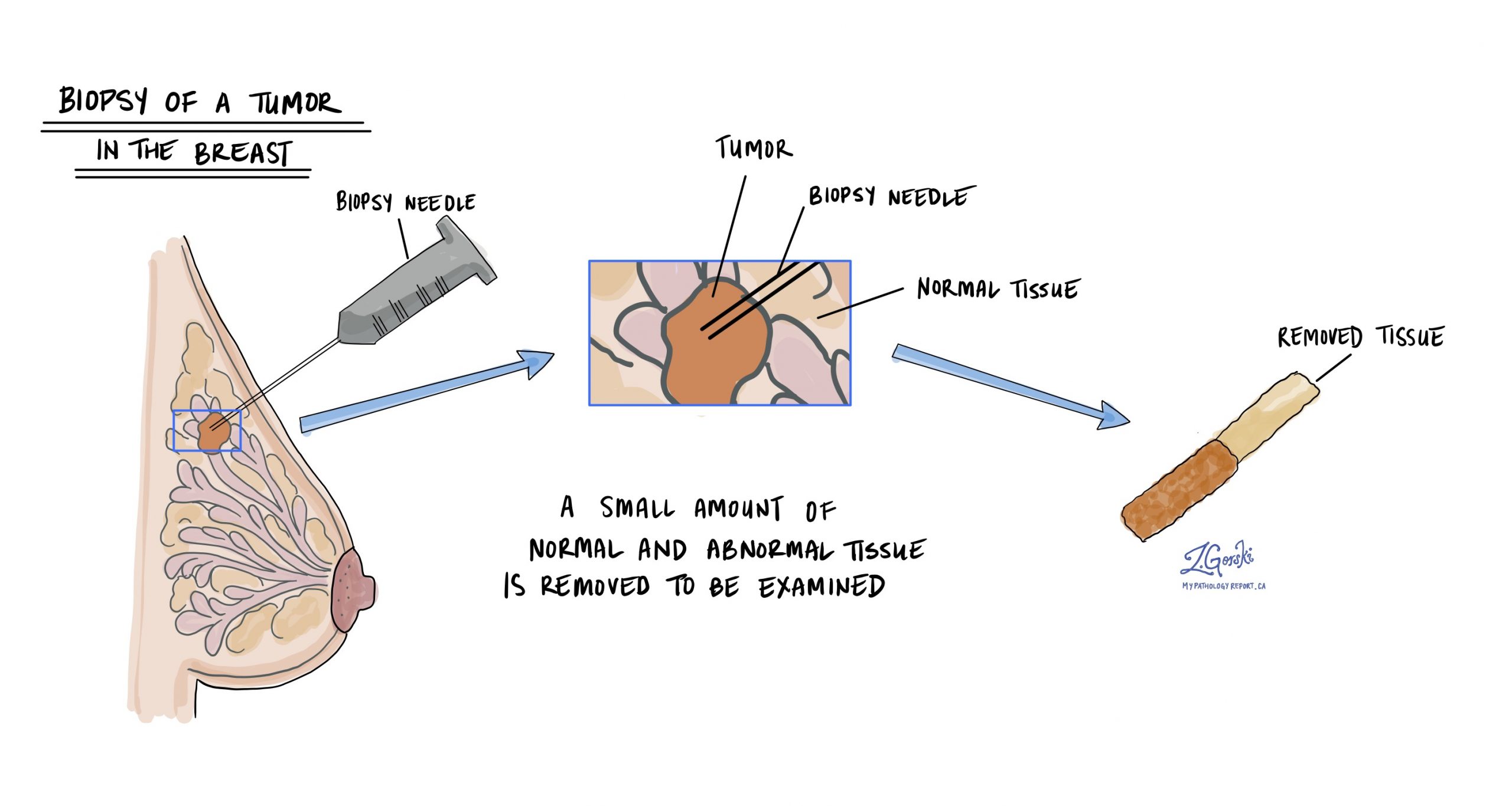
How is this diagnosis made?
The diagnosis of invasive apocrine carcinoma is usually made after a small sample of the tumour is removed in a procedure called a biopsy. The tissue is then sent to a pathologist for examination under a microscope.
Nottingham histologic grade
The Nottingham histologic grade, also known as the modified Scarff-Bloom-Richardson grade, is a system used by pathologists to evaluate breast cancer under the microscope. It helps determine the aggressiveness of the tumour and provides important information for planning treatment. The grade is based on how different the cancer cells look from normal breast cells and how quickly they are growing.
To calculate the grade, pathologists examine three features of the cancer:
- Tubule formation: This measures the extent to which cancer cells form structures resembling normal breast glands. If most of the cells form tubules, the tumour gets a lower score. Fewer tubules mean a higher score.
- Nuclear pleomorphism: This describes the variation in the appearance of cancer cells’ nuclei (the part of the cell that contains DNA) compared to normal cells. The score is low if the nuclei are uniform and similar to those of normal cells. If they are very different and irregular, the score is higher.
- Mitotic count: This measures the number of cancer cells that are actively dividing. Cells that are dividing undergo a process called mitosis and are referred to as mitotic figures. A higher number of dividing cells indicates that the tumour is growing quickly, resulting in a higher score.
Each feature is scored from 1 to 3, with 1 indicating a level close to normal and 3 indicating a more abnormal level. The scores are added together to give a total score between 3 and 9, which determines the grade.

The total score places the tumour into one of three grades:
- Grade 1 (Low grade): Total score of 3 to 5. Cancer cells often resemble normal cells and typically grow at a slow rate.
- Grade 2 (Intermediate grade): Total score of 6 to 7. The cancer cells show more differences from normal and grow at a moderate rate.
- Grade 3 (High grade): Total score of 8 to 9. Cancer cells appear distinctly different from normal cells and tend to grow more rapidly.
The grade helps doctors predict how aggressive the cancer will likely be. Grade 1 cancers often grow slowly and may have a better outcome. Grade 3 cancers can grow and spread more quickly and may require more aggressive treatment. Your doctor will use the grade and other factors, such as tumour size and whether cancer is found in lymph nodes, to guide treatment decisions.
Estrogen receptor (ER) and progesterone receptor (PR)
Hormone receptors are proteins found in some breast cancer cells. The two main types tested are estrogen receptor (ER) and progesterone receptor (PR). Cancer cells with these receptors utilise hormones such as estrogen and progesterone to promote growth and division. Testing for ER and PR helps guide treatment and predict prognosis.
Cancer cells are described as hormone receptor-positive if ER or PR is present in at least 1% of cells. These cancers often grow more slowly, are less aggressive, and typically respond well to hormone-blocking therapies, such as tamoxifen or aromatase inhibitors (e.g., anastrozole, letrozole, or exemestane). Hormone therapy helps reduce the chance of cancer recurrence.
Your pathology report will typically include:
-
Percentage of positive cells: For example, “80% ER-positive” means 80% of cancer cells have estrogen receptors.
-
Intensity of staining: Reported as weak, moderate, or strong, this indicates the number of receptors present in the cancer cells.
-
Overall score (Allred or H-score): This combines percentage and intensity, with higher scores indicating a better response to hormone therapy.
Tumours with ER positivity between 1% and 10% are considered ER low positive. These cancers still usually respond better to hormone therapy compared to ER-negative cancers.
Understanding ER and PR status helps your doctors plan effective treatment tailored to your cancer.
HER2
HER2 (human epidermal growth factor receptor 2) is a protein found on specific breast cancer cells that facilitates their growth and division. Breast cancers with extra HER2 proteins due to a change (amplification) in the HER2 gene are called HER2-positive.
HER2-positive cancers tend to be more aggressive and were once associated with a poorer prognosis. However, effective targeted therapies now significantly improve outcomes for patients with HER2-positive cancers. Knowing the HER2 status helps your doctors choose treatments specifically designed for your type of cancer, often including targeted drugs along with chemotherapy.
Two tests are commonly performed to measure HER2 in breast cancer cells: immunohistochemistry (IHC) and fluorescence in situ hybridization (FISH).
Immunohistochemistry (IHC) for HER2
Immunohistochemistry (IHC) is a test pathologists use to measure the amount of HER2 protein on the surface of breast cancer cells. To perform this test, pathologists use a small tissue sample from the tumour. They apply special antibodies to the tissue, which bind to HER2 proteins if they are present. These antibodies are then made visible under a microscope by adding a colored dye. By examining the intensity (strength) and amount of colour present, the pathologist determines how much HER2 protein is on the cancer cells.
Your pathology report will describe the results of HER2 IHC testing as a score ranging from 0 to 3+:
-
0 (negative): No visible staining, meaning no significant HER2 protein is detected. This indicates a HER2-negative tumour, and targeted HER2 treatments will not typically be helpful.
-
1+ (negative): Weak and incomplete staining. These tumours are still considered HER2-negative and usually don’t benefit from HER2-targeted treatments.
-
2+ (borderline or equivocal): Moderate staining, meaning the result is unclear. Additional testing, typically a FISH test, is required to determine whether the cancer is HER2-positive or HER2-negative.
-
3+ (positive): Strong and complete staining on the surface of the cancer cells. This indicates a HER2-positive breast cancer. HER2-positive cancers often grow faster but respond very well to HER2-targeted therapies like trastuzumab.
Fluorescence in situ hybridization (FISH) for HER2
Fluorescence in situ hybridization (FISH) is a test used to examine cancer cells for extra copies of specific genes, such as HER2. In breast cancer testing, FISH is usually performed after the initial HER2 IHC test gives unclear or borderline results.
To perform a FISH test, pathologists use a small tissue sample from the tumour. They add special fluorescent probes to the tissue, which attach specifically to the HER2 genes inside the cancer cells. Under a microscope, these probes glow brightly, enabling pathologists to count the number of copies of the HER2 gene present in each cell.
Your pathology report will typically describe the results of the FISH test as either:
-
Positive (Amplified): The cancer cells have extra copies of the HER2 gene. This is known as HER2-positive breast cancer. These cancers often grow more aggressively but typically respond well to targeted HER2 treatments, such as trastuzumab (Herceptin).
-
Negative (Non-amplified): The cancer cells have a normal number of HER2 gene copies. This is called HER2-negative breast cancer, meaning targeted HER2 therapies are usually not helpful.
Sometimes the report may describe the exact number of gene copies per cell (for example, an average HER2 copy number or a HER2-to-chromosome ratio). These detailed numbers help pathologists and oncologists accurately confirm the HER2 status, guiding the choice of the most effective treatment for your specific cancer type.
Androgen receptor (AR)
Androgen receptors (AR) are proteins found in some breast cancer cells, particularly invasive apocrine carcinoma of the breast. Cancer cells with androgen receptors use hormones such as testosterone or other androgens to help them grow and multiply. Testing for AR helps your medical team understand the biology of your tumour, guide treatment decisions, and predict how the cancer may behave.
Cancer cells are described as androgen receptor-positive if AR is present in at least 1% of the cells. Apocrine breast cancers often have strong and widespread AR positivity, typically greater than 10%. Tumours with a high percentage of AR-positive cells may respond to targeted therapies that block androgen signalling, such as bicalutamide or enzalutamide. These treatments may help slow the growth of the cancer and reduce the risk of recurrence.
Your pathology report will typically include:
- Percentage of positive cells: For example, “90% AR-positive” means that 90% of the cancer cells have androgen receptors.
- Intensity of staining: Reported as weak, moderate, or strong, this indicates how many androgen receptors are present in the cancer cells.
- Overall assessment: Often described by combining percentage and intensity to evaluate the likelihood of response to targeted therapies.
Understanding the AR status of your cancer helps your healthcare team choose the most effective treatments tailored specifically to your diagnosis.
Tumour size
The size of a breast tumour is important because it is used to determine the pathologic tumour stage (pT) and because larger tumours are more likely to metastasize (spread) to lymph nodes and other parts of the body. The tumour size can only be determined after the entire tumour has been removed. For this reason, it will not be included in your pathology report after a biopsy.
Tumour extension
Invasive apocrine carcinoma starts inside the breast, but the tumour may spread into the overlying skin or the muscles of the chest wall. The term tumour extension is used when tumour cells are found in the skin or muscles below the breast. Tumour extension is important because it is associated with a higher risk that the tumour will recur after treatment (local recurrence) or that cancer cells will metastasise to a distant body site, such as the lungs. It is also used to determine the pathologic tumour stage (pT).
Lymphovascular invasion
Lymphovascular invasion (LVI) means that cancer cells have entered small blood vessels or lymphatic channels located near the tumour in the breast. Blood vessels are small tubes that carry blood to and from tissues, delivering oxygen and nutrients. Lymphatic vessels are similar but carry lymph fluid, which contains immune cells and helps remove waste from tissues. Both types of vessels can provide pathways for cancer cells to spread beyond the original tumour site.
Pathologists carefully examine tissue samples under the microscope to look for LVI. Cancer cells may appear as single cells or clusters inside these vessels, often surrounded by a clear space that marks the vessel wall. Your pathology report will typically describe LVI as positive (or present) if cancer cells are found inside these vessels, or negative (or absent) if no cancer cells are seen. The presence of LVI is important because it suggests a higher chance that cancer will spread to nearby lymph nodes or may spread to distant parts of the body. Because of this, your medical team may recommend more aggressive treatments, such as chemotherapy, radiation therapy, or targeted therapy, to reduce the risk of cancer recurrence and spread.

Margins
In pathology, a margin is the edge or border of tissue removed along with a tumour during surgery. Margins are closely examined under a microscope by a pathologist to see if any cancer cells are present at the cut edge. The status of these margins is important because it helps determine if the entire tumour was removed or if cancer cells may have been left behind in the body.
Margins are typically assessed only after a surgical procedure, such as a resection or excision, which removes the entire tumour. They are usually not described following a biopsy, as biopsies remove only a small sample of tissue, not the whole tumour. The number and type of margins described in your pathology report depend on the size and location of the tumour, as well as the type of tissue removed.
To evaluate margins, the pathologist carefully examines thin slices of the tissue under a microscope. They look closely at the edges to see if tumour cells reach the cut surface. Your pathology report will describe these results as either negative (no cancer cells seen at the margin) or positive (cancer cells present at the margin). If the margin is negative, the report may also mention the exact distance between the closest tumour cells and the cut edge, known as the margin width.
The results of the margin examination are very important for planning your treatment. A positive margin indicates that some cancer cells are likely to remain in the body, thereby increasing the risk of the cancer recurring or progressing. If you have a positive margin, your doctor may recommend further treatment, such as additional surgery to remove any remaining tumour or radiation therapy directed at the area where the positive margin was found. A negative margin, especially with a greater distance from tumour cells to the cut edge, suggests that the cancer has been entirely removed, reducing the likelihood of recurrence.

Lymph nodes
Lymph nodes are small, bean-shaped organs that play a key role in the body’s immune system. They filter harmful substances, such as bacteria, viruses, and cancer cells, from the lymph fluid circulating throughout the body. Lymph nodes contain immune cells that help fight infections and can respond to cancer cells.
When breast cancer spreads beyond the original tumour, it often moves first into nearby lymph nodes. Because of this, pathologists carefully examine lymph nodes removed during surgery to determine whether cancer has spread. This is a critical step for understanding your cancer’s stage, planning appropriate treatment, and predicting your prognosis (the likely outcome).
In breast cancer, several types of lymph nodes may be examined:
-
Axillary lymph nodes: Located under the arm, these are the most commonly examined nodes in breast cancer cases.
-
Sentinel lymph nodes: These are usually the first few lymph nodes that cancer cells would reach from the breast. A sentinel lymph node biopsy involves removing one or a few of these nodes for testing.
-
Internal mammary lymph nodes: Located near the breastbone, these nodes are sometimes examined if cancer cells are found in other lymph nodes or if imaging tests suggest involvement.

How lymph nodes are examined
Lymph nodes removed during surgery are carefully examined under a microscope by a pathologist.
Your pathology report will provide detailed information, including:
-
Number of lymph nodes examined: The total count of lymph nodes removed and examined.
-
Number of positive lymph nodes: The number of lymph nodes containing cancer cells.
-
Size of cancer deposits: The size of the largest group of cancer cells found within any lymph node.
-
Other features: Your report may occasionally mention additional findings, such as extranodal extension, which indicates that cancer cells have spread beyond the lymph node itself.
Possible results and what they mean
Your pathology report may describe cancer cells in the lymph nodes using the following terms, depending on the size of the cancer deposits:
-
Isolated tumour cells (ITCs): Very small groups of cancer cells measuring 0.2 millimeters or less. Although these cells are detected, lymph nodes containing only isolated tumour cells are generally not considered positive for the purposes of cancer staging, and they usually have minimal impact on prognosis and treatment decisions.
-
Micrometastasis: Cancer cell clusters that measure between 0.2 millimeters and 2 millimeters. If lymph nodes contain only micrometastases, this finding is noted as “pN1mi” in your report. Micrometastases can slightly increase the risk of recurrence and may lead your doctors to consider additional treatments, such as radiation or chemotherapy, depending on other factors.
-
Macrometastasis: Larger cancer cell clusters measuring more than 2 millimeters. The presence of macrometastases indicates a greater risk that the cancer may recur or spread further. These findings typically prompt more aggressive treatment strategies, which can include additional surgery, chemotherapy, radiation therapy, or targeted therapies.

Residual cancer burden index
The residual cancer burden (RCB) index measures the amount of cancer remaining in the breast and nearby lymph nodes after neoadjuvant therapy (treatment given before surgery). The index combines several pathologic features into a single score and classifies the cancer’s response to treatment. Doctors at the University of Texas MD Anderson Cancer Center developed the RCB (http://www.mdanderson.org/breastcancer_RCB).
Here’s how the score is calculated:
- Size of the tumour bed in the breast: Pathologists measure the largest two dimensions of the area where the tumour was located, called the tumour bed. This area may contain a mix of normal tissue, cancer cells, and scar tissue from the therapy.
- Cancer cellularity: Cancer cellularity estimates the percentage of the tumour bed that still contains cancer cells. This includes both invasive cancer (cancer that has spread into surrounding tissue) and in situ cancer (cancer cells that have not spread).
- Percentage of in situ disease: Within the tumour bed, pathologists also estimate the percentage of cancer that is in situ, meaning that the cancer cells are confined to the milk ducts or lobules and have not spread into the surrounding tissue.
- Lymph node involvement: The number of lymph nodes containing cancer cells (positive lymph nodes) is counted, and the size of the largest cluster of cancer cells in the lymph nodes is also measured.
These features are combined using a standardized formula to calculate the RCB score.
Based on the RCB score, patients are divided into four categories:
- RCB-0 (pathologic complete response): No residual invasive cancer is detected in the breast or lymph nodes.
- RCB-I (minimal burden): Very little residual cancer is present.
- RCB-II (moderate burden): A moderate amount of cancer remains.
- RCB-III (extensive burden): A large amount of cancer remains in the breast or lymph nodes.
The RCB classification helps predict a patient’s likelihood of staying cancer-free after treatment. Patients with an RCB-0 classification typically have the best outcomes, with the highest chances of long-term survival without recurrence. As the RCB category increases from RCB-I to RCB-III, the risk of cancer recurrence increases, which may prompt additional treatments to reduce this risk.
Pathologic stage for invasive apocrine carcinoma
The pathologic staging system for invasive apocrine carcinoma of the breast helps doctors understand how far the cancer has spread and plan the best treatment. The system mainly uses the TNM staging, which stands for Tumor, Nodes, and Metastasis. Early-stage cancers (like T1 or N0) might only require surgery and possibly radiation, while more advanced stages (like T3 or N3) may need a combination of surgery, radiation, chemotherapy, and targeted therapies. Proper staging ensures that patients receive the most effective treatments based on the extent of their disease, which can improve survival rates and quality of life.
Tumour stage (pT)
This feature examines the size and extent of the breast tumour. The tumour is measured in centimetres, and its growth beyond the breast tissue is assessed.
T0: No evidence of primary tumour. This means no tumour can be found in the breast.
T1: The tumour is 2 centimetres or smaller in greatest dimension. This stage is further subdivided into:
- T1mi: Tumour is 1 millimetre or smaller.
- T1a: Tumour is larger than 1 millimetre but not larger than 5 millimetres.
- T1b: Tumour is larger than 5 millimetres but not larger than 10 millimetres.
- T1c: Tumour is larger than 10 millimetres but not over 20 millimetres.
T2: The tumour is larger than 2 centimetres but not larger than 5 centimetres.
T3: The tumour is larger than 5 centimetres.
T4: The tumour has spread to the chest wall or skin, regardless of its size. This stage is further subdivided into:
- T4a: Tumour has invaded the chest wall.
- T4b: Tumour has spread to the skin, causing ulcers or swelling.
- T4c: Both T4a and T4b are present.
- T4d: Inflammatory breast cancer, characterized by redness and swelling of the breast skin.
Nodal stage (pN)
This feature examines if the cancer has spread to the nearby lymph nodes, which are small, bean-shaped structures found throughout the body.
N0: No cancer is found in the nearby lymph nodes.
N0(i+): Isolated tumour cells only.
N1: Cancer has spread to 1 to 3 axillary lymph nodes (under the arm).
- N1mi: Micrometastases only.
- N1a: Metastases in 1-3 axillary lymph nodes, at least one metastasis larger than 2.0 mm.
- N1b: Metastases in ipsilateral internal mammary sentinel nodes, excluding ITCs
N2: Cancer has spread to:
- N2a: 4 to 9 axillary lymph nodes.
- N2b: Internal mammary lymph nodes without involvement of axillary lymph nodes.
N3: Cancer has spread to:
- N3a: 10 or more axillary lymph nodes or two infraclavicular lymph nodes (below the collarbone).
- N3b: Internal mammary lymph nodes and axillary lymph nodes.
- N3c: Supraclavicular lymph nodes (above the collarbone).
Other helpful resources
American Breast Cancer Foundation
Canadian Breast Cancer Foundation
We are proud to partner with:
![]()