by Jason Wasserman MD PhD FRCPC
November 30, 2024
Lymphoepithelial carcinoma is a rare type of cancer that typically develops in the head and neck area, most often in the salivary glands. It is characterized by a mix of cancerous cells and a large number of immune cells called lymphocytes. This type of cancer is considered aggressive but can often be treated successfully if diagnosed early.

What are the symptoms of lymphoepithelial carcinoma?
The symptoms of lymphoepithelial carcinoma depend on where the tumour develops. Common symptoms include a painless lump in the neck or near the jaw, difficulty swallowing, persistent ear pain, or voice changes. Some people may also notice swelling in the salivary glands or other head and neck areas.
What causes lymphoepithelial carcinoma?
The exact cause of lymphoepithelial carcinoma is not fully understood. However, it has been linked to the Epstein-Barr virus (EBV), especially in specific populations and regions of the world. Genetic and environmental factors may also play a role in its development.
How is this diagnosis made?
The diagnosis of lymphoepithelial carcinoma is made by examining a tissue sample under a microscope. The tissue is usually taken through a biopsy, which involves removing a small piece of the tumour. Pathologists use special techniques, including stains and molecular tests, to confirm the diagnosis and rule out other types of cancer.
What are the microscopic features of lymphoepithelial carcinoma?
Under the microscope, lymphoepithelial carcinoma appears similar to a type of cancer called non-keratinizing nasopharyngeal carcinoma. The tumour consists of nests of large cancer cells surrounded by a dense background of immune cells called lymphocytes. The tissue around the tumour often shows inflammation, and in the salivary glands, this may look like a condition called lymphoepithelial sialadenitis, a type of chronic immune reaction.

What other tests may be performed to confirm the diagnosis?
Pathologists often perform a test called in situ hybridization to confirm the diagnosis. This test detects small pieces of genetic material from the Epstein-Barr virus (EBV) in the tumour cells. EBER (Epstein-Barr virus-encoded RNA) is a common version of this test. If the tumour cells show evidence of EBV, it supports the diagnosis of lymphoepithelial carcinoma, particularly in populations where this virus is known to play a role in cancer development.
Extraparenchymal extension
In the context of a salivary gland tumour such as lymphoepithelial carcinoma, extraparenchymal extension (EPE) is the spread of the tumour beyond the salivary gland into the surrounding tissues. This condition is often associated with a more aggressive form of cancer, indicating that the tumour can invade beyond its original site. The presence of extraparenchymal extension is associated with more aggressive tumours and a worse prognosis.
Extraparenchyma, extension impacts the pathologic stage but only for tumours arising from one of the major salivary glands (parotid, submandibular, and sublingual). Tumours with extraparenchymal extension are generally classified at a higher stage, reflecting their advanced nature and the associated challenges in treatment and management.
Lymphovascular invasion
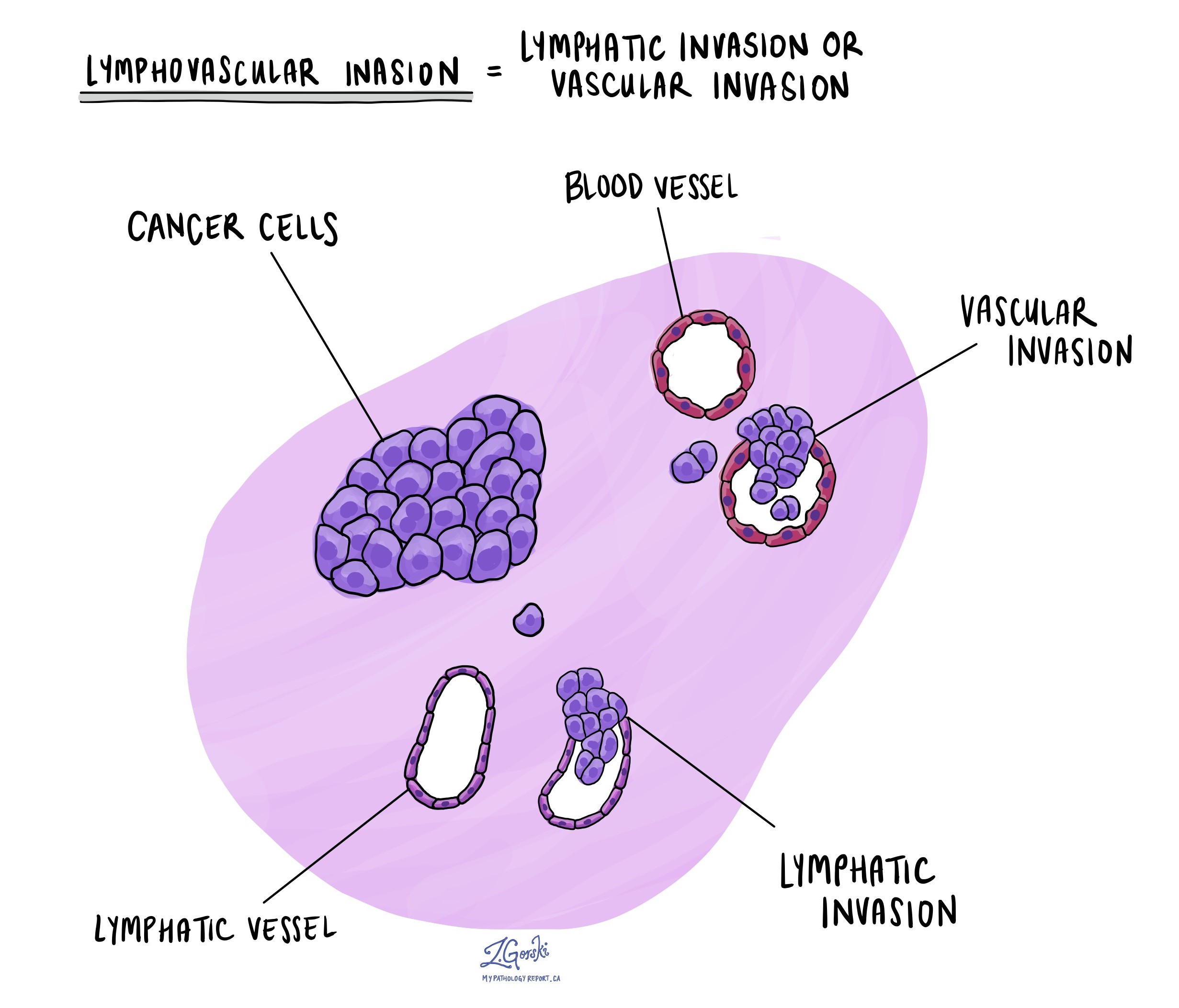
Lymphovascular invasion occurs when cancer cells invade a blood vessel or lymphatic vessel. Blood vessels are thin tubes that carry blood throughout the body, unlike lymphatic vessels, which carry a fluid called lymph instead of blood. These lymphatic vessels connect to small immune organs known as lymph nodes scattered throughout the body. Lymphovascular invasion is important because it spreads cancer cells to other body parts, including lymph nodes or the liver, via the blood or lymphatic vessels.

Perineural invasion
Pathologists use the term “perineural invasion” to describe a situation where cancer cells attach to or invade a nerve. “Intraneural invasion” is a related term that specifically refers to cancer cells inside a nerve. Nerves, resembling long wires, consist of groups of cells known as neurons. These nerves, present throughout the body, transmit information such as temperature, pressure, and pain between the body and the brain. Perineural invasion is important because it allows cancer cells to travel along the nerve into nearby organs and tissues, raising the risk of the tumour recurring after surgery.

Margins
In pathology, a margin is the edge of tissue removed during tumour surgery. The margin status in a pathology report is important as it indicates whether the entire tumour was removed or if some was left behind. This information helps determine the need for further treatment.
Pathologists typically assess margins following a surgical procedure, like an excision or resection, that removes the entire tumour. Margins aren’t usually evaluated after a biopsy, which removes only part of the tumour. The number of margins reported and their size—how much normal tissue is between the tumour and the cut edge—vary based on the tissue type and tumour location.
Pathologists examine margins to check if tumour cells are at the tissue’s cut edge. A positive margin, where tumour cells are found, suggests that some cancer may remain in the body. In contrast, a negative margin, with no tumour cells at the edge, suggests the tumour was fully removed. Some reports also measure the distance between the nearest tumour cells and the margin, even if all margins are negative.

Lymph nodes
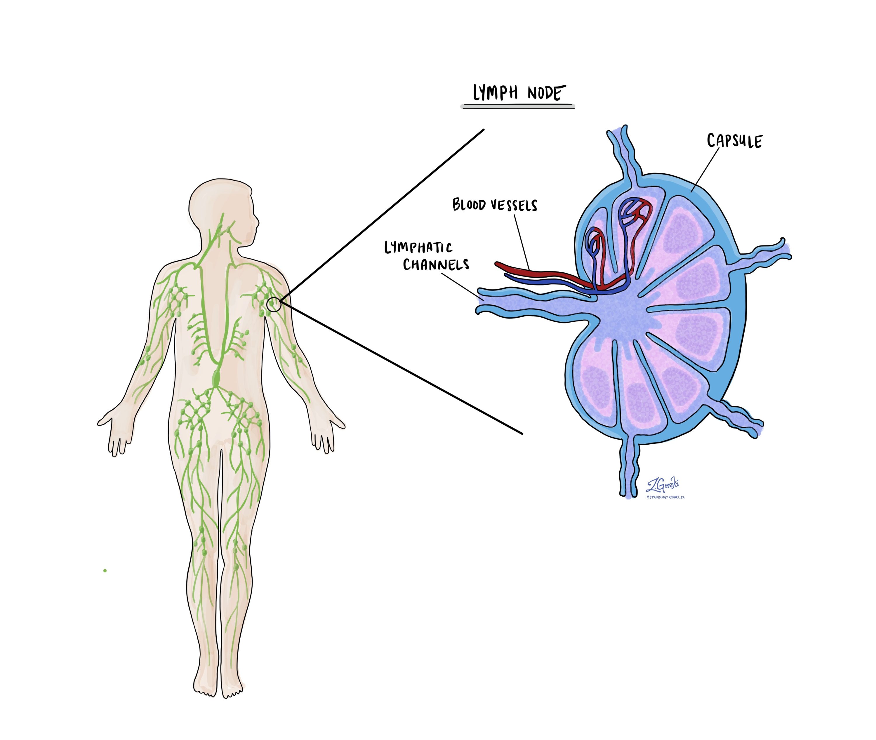
Small immune organs, known as lymph nodes, are located throughout the body. Cancer cells can travel from a tumour to these lymph nodes via tiny lymphatic vessels. For this reason, doctors often remove and microscopically examine lymph nodes to look for cancer cells. This process, where cancer cells move from the original tumour to another body part, like a lymph node, is termed metastasis.
Cancer cells usually first migrate to lymph nodes near the tumour, although distant lymph nodes may also be affected. Consequently, surgeons typically remove lymph nodes closest to the tumour first. They might remove lymph nodes farther from the tumour if they are enlarged and there’s a strong suspicion they contain cancer cells.

Pathologists will examine any lymph nodes removed under a microscope, and the findings will be detailed in your report. A “positive” result indicates the presence of cancer cells in the lymph node, while a “negative” result means no cancer cells were found. If the report finds cancer cells in a lymph node, it might also specify the size of the largest cluster of these cells, often referred to as a “focus” or “deposit.” Extranodal extension occurs when tumour cells penetrate the lymph node’s outer capsule and spread into the adjacent tissue.
Examining lymph nodes is important for two reasons. First, it helps determine the pathologic nodal stage (pN). Second, discovering cancer cells in a lymph node suggests an increased risk of later finding cancer cells in other body parts. This information guides your doctor in deciding whether you need additional treatments, such as chemotherapy, radiation therapy, or immunotherapy.
Pathologic stage
Pathologic staging is a system doctors use to describe the size and spread of a tumour. This helps determine how advanced the cancer is and guides treatment decisions. The pathologic stage is usually determined after the tumour is removed and examined by a pathologist, who analyzes the tissue under a microscope. For acinic cell carcinoma, staging is based on the “TNM” system, where “T” stands for the size and extent of the primary tumour, “N” refers to lymph node involvement, and “M” indicates whether the cancer has spread to other parts of the body.
Tumour stage (pT)
The tumour stage describes the size of the tumour in the salivary gland and whether it has spread into nearby tissues.
- T0 means there is no evidence of a primary tumour in the salivary gland.
- Tis refers to carcinoma “in situ,” meaning the cancer cells are limited to where they started and have not invaded deeper tissues.
- T1 means the tumour is 2 cm or smaller and has not spread beyond the salivary gland.
- T2 refers to a tumour larger than 2 cm but not larger than 4 cm, still confined to the salivary gland.
- T3 means the tumour is larger than 4 cm or has spread to nearby soft tissues.
- T4 describes more advanced tumours. T4a means the tumour has spread to the skin, jawbone, ear canal, or facial nerve. T4b indicates very advanced cancer that has spread to the base of the skull, nearby bones, or major blood vessels.
Nodal stage (pN)
The nodal stage indicates whether the cancer has spread to the lymph nodes, which are small glands that help the body fight infection. Lymph node involvement can increase the risk of cancer spreading further.
- Nx means that no lymph nodes were submitted for examination.
- N0 means there is no spread to nearby lymph nodes.
- N1 indicates the cancer has spread to a single lymph node on the same side of the neck, measuring 3 cm or smaller.
- N2 describes more extensive lymph node involvement:
- N2a: A single lymph node on the same side of the neck is affected, measuring up to 6 cm, or smaller nodes that show signs of cancer outside the node.
- N2b: Multiple lymph nodes on the same side of the neck are affected, none larger than 6 cm.
- N2c: Cancer has spread to lymph nodes on both sides of the neck or on the opposite side, none larger than 6 cm.
- N3 indicates more advanced lymph node involvement. N3a means a node larger than 6 cm is affected. N3b involves multiple nodes or any nodes where cancer has spread outside the lymph node into nearby tissues.
What is the prognosis for a person diagnosed with lymphoepithelial carcinoma?
The outlook for people with lymphoepithelial carcinoma is generally good, with a five-year survival rate of about 81%. The presence of cancer in nearby lymph nodes is seen in about 17% of cases, while the spread of cancer to distant parts of the body is uncommon, occurring in about 6% of cases. In areas of the world where this cancer is more common, distant spread may happen more frequently. The involvement of lymph nodes or distant organs is important in determining the prognosis. Early detection and treatment play a critical role in achieving the best outcomes.


