
by Jason Wasserman MD PhD FRCPC
July 16, 2024
Background:
Moderate keratinizing squamous dysplasia is a precancerous condition characterized by abnormal growth and organization of squamous cells in the epithelium of the oral cavity. In this condition, the squamous cells exhibit atypia (abnormalities in size, shape, and organization) and increased keratin production, but the changes are less extensive than in severe dysplasia. The dysplasia is classified as moderate when these cellular abnormalities involve most but not all of the epithelial thickness.
What parts of the oral cavity typically involve moderate keratinizing squamous dysplasia?
Moderate keratinizing squamous dysplasia can occur in various parts of the oral cavity, including:
- Tongue: Particularly the lateral borders and ventral surface.
- Floor of the mouth: An area under the tongue.
- Buccal mucosa: The inner lining of the cheeks.
- Gingiva: The gums.
- Hard palate: The roof of the mouth.
- Lips: The inner surface.
What are the symptoms of moderate keratinizing squamous dysplasia?
The symptoms of moderate keratinizing squamous dysplasia can be subtle and may include:
- White or red patches (leukoplakia or erythroplakia): These are areas of abnormal tissue that can be seen on the mucosal surfaces.
- Pain: Affected areas may be painful or sensitive.
- Thickened or rough areas: The surface may feel rough or thickened due to increased keratin production.
- Difficulty in chewing or swallowing: If the dysplasia is extensive, it may interfere with normal oral functions such as chewing or swallowing.
- Numbness: There may be a sensation of numbness in the affected areas.
What causes moderate keratinizing squamous dysplasia?
Several factors can contribute to the development of moderate keratinizing squamous dysplasia, including:
- Tobacco use: Both smoking and smokeless tobacco are major risk factors.
- Alcohol consumption: Heavy alcohol use can exacerbate the effects of tobacco and independently increase risk.
- Chronic inflammatory conditions: Conditions like lichen planus can lead to chronic inflammation and increase the risk of dysplasia.
- Immune suppression/immune deficiency: Conditions or treatments that suppress the immune system can increase the risk of dysplastic changes due to reduced immune surveillance.
- Betel nut chewing: Chewing betel nut (areca nut) is a significant risk factor, particularly in regions where this practice is common. Betel nut contains carcinogenic compounds that can lead to oral mucosal changes and dysplasia.
Is moderate keratinizing squamous dysplasia associated with an increased risk of developing cancer of the oral cavity?
Yes, moderate keratinizing squamous dysplasia is associated with an increased risk of developing squamous cell carcinoma, a type of oral cavity cancer. The risk is lower than that associated with severe dysplasia but higher than with mild dysplasia. Therefore, early detection and appropriate management of dysplastic lesions are important in preventing the progression to oral cancer.
What are the microscopic features of moderate keratinizing squamous dysplasia?
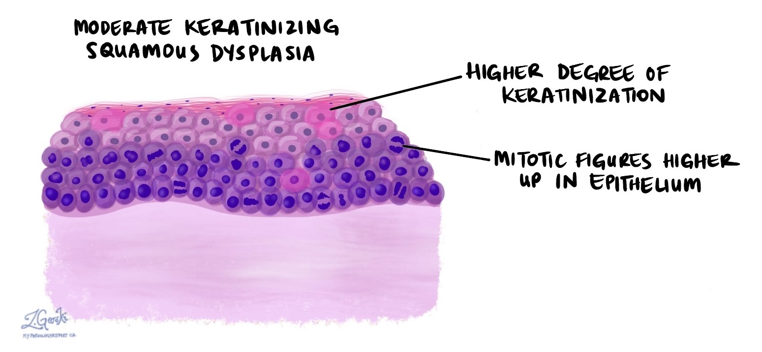
Microscopically, moderate keratinizing squamous dysplasia shows a disorganized epithelial structure with loss of normal stratification and architecture, but these changes are typically confined to the lower two-thirds of the epithelium. The cells display abnormalities in size, shape, and nuclear features, including hyperchromasia (darkly staining nuclei), pleomorphism (variation in cell and nuclear size and shape), and an increased nuclear-to-cytoplasmic ratio. There is an increased number of mitotic figures, but they are usually confined to the lower layers of the epithelium. The surface is typically thickened and keratotic. Additionally, there may be chronic inflammation in the underlying connective tissue, which can accompany the dysplastic changes.
Margins
A margin is any tissue that the surgeon cuts to remove the abnormal tissue area from your body. The types of margins described in your report will depend on the area of the oral cavity involved and the type of surgery performed. Margins are usually only described in your report after the entire abnormal tissue area has been removed.
A negative margin means that dysplasia was not seen at any of the cut edges of the tissue. A margin is called positive when dysplasia is seen at the very edge of the cut tissue. A positive margin is associated with a higher risk that dysplasia will come back at the same site after treatment.

About this article
Doctors wrote this article to help you read and understand your pathology report. If you have any questions, please contact us.
Learn more pathology
Atlas of Pathology
We are proud to partner with:
![]()
Our work is generously supported by:
![]()