by Rosemarie Tremblay-LeMay MD FRCPC
May 1, 2024
Myelodysplastic syndrome (MDS) is a group of diseases where new blood cells produced in the bone marrow are abnormal and do not function properly. MDS increases the risk of developing acute myeloid leukemia, a type of blood cancer.
What cells are normally found in the blood?
Normal blood is made up of many different kinds of cells. For example, it contains red blood cells that carry oxygen from our lungs to our body and carbon dioxide from our body back to our lungs. It also contains specialized immune cells, including neutrophils, lymphocytes, and monocytes, that protect us against infections and help our body heal after an injury. Finally, the blood contains platelets that help stop bleeding after an injury by creating a blood clot.
Most new blood cells are made in the marrow, a part of the bone that is damaged by disease. This process is called hematopoiesis. Because hematopoiesis takes place in the marrow, any disease that damages it can decrease or prevent the production of new blood cells.
What medical conditions are associated with myelodysplastic syndrome?
Patients with MDS may experience a variety of medical issues depending on the types of cells that are abnormal.
The most common conditions of MDS include:
- Anemia – Anemia is caused by a decreased number of normal red blood cells. People with anemia may experience shortness of breath, chest pain, and decreased energy.
- Neutropenia – Neutropenia is caused by a decreased number of specialized immune cells called neutrophils. People with neutropenia are more likely to develop infections.
- Thrombocytopenia – Thrombocytopenia is caused by a decreased number of specialized blood cells called platelets. People with thrombocytopenia are unable to form blood clots and may bleed excessively after an injury or have spontaneous bleeding.
- Pancytopenia – Doctors use the term pancytopenia when there is a decreased number of red blood cells, neutrophils, and platelets. People with pancytopenia may experience all of the complications listed above.
What causes myelodysplastic syndrome?
Within the bone marrow, there are a small number of specialized immature cells that multiply to produce new mature blood cells. MDS is caused by a genetic change in one of these immature bone marrow cells. As the abnormal cell multiplies, all of the new cells created will have the same genetic change. Eventually, these abnormal cells replace all of the normal, healthy cells in the bone marrow.
Throughout this process, new genetic changes can develop. These new changes can cause the disease to become more aggressive and less likely to respond to therapy. For example, acute myeloid leukemia is a type of blood cancer that can develop from MDS over time.
How is this diagnosis made?
The diagnosis of MDS is usually made after a small sample of bone marrow is removed in a procedure called biopsy and aspiration. A pathologist then examines the sample under a microscope. An additional test called a karyotype may also be performed to look for changes in the genetic material or chromosomes of each cell.
Microscopic features of myelodysplastic syndrome
When examining the bone marrow sample under the microscope, your pathologist will look for each type of blood cell normally found in the bone marrow to ensure that hematopoiesis is taking place normally. Immature cells go through a sequence of stages until they become a type of mature blood cell. All cells in the process of maturing to become a given type of blood cell are called a lineage.
Dysplasia
If MDS is present, the blood cells in the bone marrow will look abnormal in shape, size, or colour. Pathologists call this change dysplasia. To diagnose MDS, your pathologist must see dysplasia affecting at least 10% of the blood cells of one type or lineage. Single-lineage dysplasia means that dysplasia affects only one type of blood cell. Multilineage dysplasia means that dysplasia affects more than one type of blood cell.
Karyotype
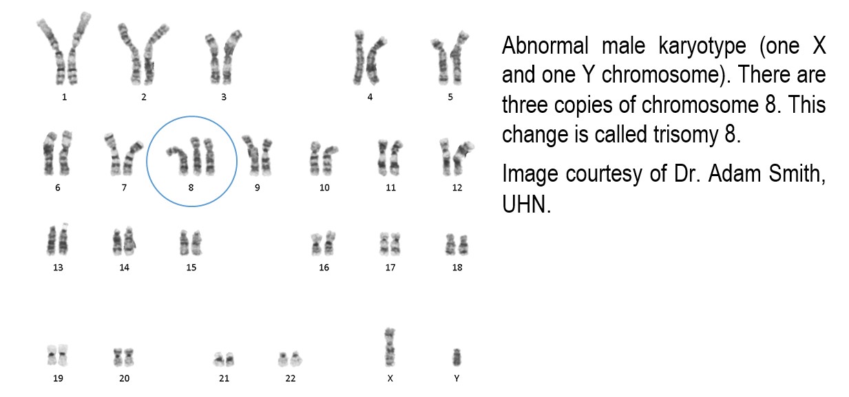
A karyotype is a test where the chromosomes, which contain your DNA, are stained with a special dye so that they can be examined under a microscope. Normal cells contain 23 pairs of chromosomes.

Abnormalities that can be seen on a karyotype test include a gain or loss of a chromosome, loss of a piece of the chromosome, or an exchange of genetic material between chromosomes. A complex karyotype means that three or more of these abnormalities were found.

Not all cases of MDS will have an abnormal karyotype. When the karyotype is abnormal, all the daughter cells that came from the original abnormal cell will share the same abnormalities. This entire group of cells is called a clone.
Some abnormalities are associated with better outcomes and a better response to treatment, while others are associated with worse outcomes and are less responsive to treatment. For this reason, the karyotype is an important part of the evaluation of MDS.
Some abnormalities in the karyotype are rarely seen in healthy individuals. These abnormalities allow your doctor to make the diagnosis of MDS even if dysplasia is not seen or it is seen affecting less than 10% of cells. In this case, the disease would be called MDS unclassifiable.
Finally, some genetic changes involved in the development of MDS are too small to be seen by the karyotype test. To look for small changes, your doctor may perform an additional test called next-generation sequencing.
Types of myelodysplastic syndromes
Myelodysplastic syndromes are divided into types based on the number of blood cell types (lineages) that show dysplasia, the number of immature cells, and the genetic changes found.
Types of MDS include:
- Myelodysplastic syndrome with single lineage dysplasia.
- Myelodysplastic syndrome with multilineage dysplasia.
- Myelodysplastic syndrome with ring sideroblasts (see Ring sideroblasts below).
- Myelodysplastic syndrome with excess blasts (see Myelodysplastic syndrome with excess blasts below).
- Myelodysplastic syndrome with isolated deletion of chromosome 5q.
Any MDS that occurs in a person who has a history of treatment with chemotherapy or radiation therapy to bone marrow will be classified as a therapy-related myeloid neoplasm. This is a distinct category of disease.
Are some forms of myelodysplastic syndrome inherited?
Most people who develop MDS have no known risk factors. Doctors describe these as sporadic. Rarely, a person will inherit a genetic change that will make them more likely to develop MDS and other diseases. This type of genetic change is called a germline mutation because it is found in all the cells of the body. In contrast, a genetic change that develops later in life is called a somatic or acquired mutation. Somatic or acquired mutations are only in some cells.
Some inherited or germline mutations are associated with physical changes that may lead the person or family to seek medical advice early in life. Some people will only have changes in their blood, like decreased numbers of blood cells, without other physical changes. For other people with a germline mutation, there will be no detectable sign until they develop MDS or another bone marrow disease.
Your doctor may perform tests to look for inherited or germline mutations if other members of your family have been diagnosed with bone marrow diseases or low blood counts.
Ring sideroblasts
The body uses iron to make normal, healthy red blood cells. When it is not being used, iron is stored in the bone marrow inside specialized cells called macrophages. A small amount of this iron is also held inside immature red blood cells. The iron inside these cells can be seen under the microscope as small dots within the body of the cell. Pathologists call these dots granules, and only a couple of dots are normally seen in healthy cells.
Ring sideroblasts are immature red blood cells that have extra iron inside the cytoplasm or body of the cell. This extra iron creates a tight ring around the nucleus of the cell. The diagnosis of MDS with ring sideroblasts is made if more than 15% of the immature red blood cells are ring sideroblasts and there are no findings that would fit better in a different type of MDS (for example, increased numbers of blasts, as discussed below). The diagnosis of MDS with ring sideroblasts can also be made when only 5% of the immature red blood cells are ring sideroblasts and if the patient is known to have a genetic change in a gene called SF3B1.
Other conditions that can cause ring sideroblasts must always be excluded. These conditions include copper deficiency, certain toxins, medications, and inherited diseases associated with ring sideroblasts.
Myelodysplastic syndrome with excess blasts
Many types of blood cells come from a specialized immature cell called a myeloblast. In pathology reports, these cells are usually described simply as blasts. A pathologist will make the diagnosis of MDS with excess blasts (MDS-EB) if the number of blasts is at least 2% in the blood or at least 5% in the bone marrow.
MDS with excess blasts is divided into two categories:
- MDS-EB1 – 2-4% blasts in the blood or 5-9% blasts in the bone marrow.
- MDS-EB2 – 5-19% blasts in the blood, 10-19% blasts in the bone marrow, or cells with Auer rods.
Blasts are important because if more than 20% of the cells in your blood or bone marrow are blasts, the diagnosis changes from MDS to acute myeloid leukemia.
Are there other conditions that can cause changes similar to myelodysplastic syndrome?
Myelodysplastic syndromes are not the only conditions that can cause bone marrow dysplasia or decreased blood counts. If you have cytopenia or dysplasia, your doctor will take a thorough history and perform a variety of tests to determine the exact cause.
Other conditions associated with dysplasia include:
- Vitamin B12 deficiency.
- Copper deficiency.
- Certain medications.
- Excess alcohol intake.
- Heavy metal poisoning.
- Certain auto-immune diseases.
- Infections.
Other types of bone marrow disease, for example, chronic myelomonocytic leukemia, can be associated with dysplasia. Your pathologist and other doctors will review your history and the findings in the bone marrow to reach the correct diagnosis.
About this article
Doctors wrote this article to help you read and understand your pathology report. Contact us if you have any questions about this article or your pathology report. Read this article for a more general introduction to the parts of a typical pathology report.


