

by Jason Wasserman MD PhD FRCPC
April 3, 2025
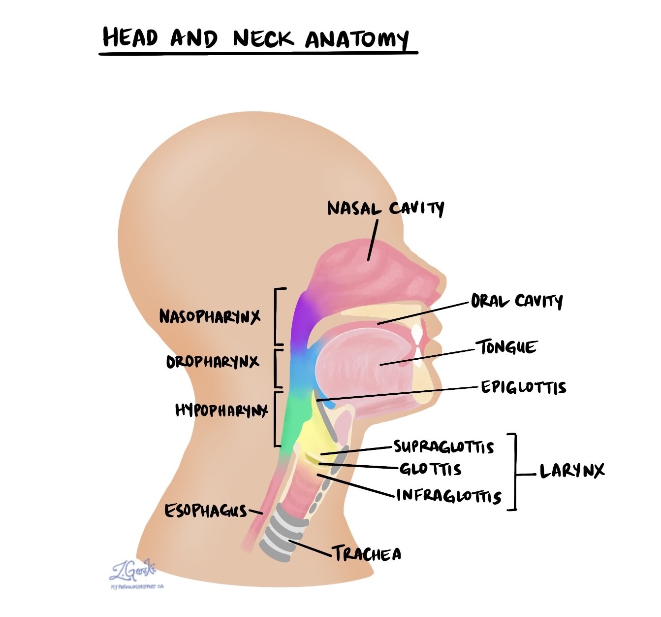
A neuroendocrine tumor (NET) of the larynx (also known as a well differentiated neuroendocrine tumor) is a rare type of cancer that develops from specialized cells called neuroendocrine cells, which release hormones into the blood in response to signals from the nervous system. These tumors occur in the larynx (voice box), the structure in your neck responsible for sound production and protecting your airway during swallowing and breathing.
What are the symptoms of a neuroendocrine tumor of the larynx?
Common symptoms of a neuroendocrine tumor in the larynx include:
- Hoarseness or persistent voice changes.
- Difficulty swallowing.
- Shortness of breath or breathing difficulties.
- Throat discomfort or pain.
Because many other conditions can also cause these symptoms, it’s important to have them evaluated by a healthcare provider.
What causes a neuroendocrine tumor of the larynx?
The exact cause of neuroendocrine tumors of the larynx is not clearly understood. Unlike the more common squamous cell carcinoma of the larynx, smoking and alcohol use are not strongly linked to these tumors. Researchers continue to study potential genetic and environmental factors, but no specific cause has yet been identified.
How is this diagnosis made?
The diagnosis of a neuroendocrine tumor is usually made by examining a biopsy (tissue sample) under a microscope. The biopsy is typically taken during a minor surgical procedure. A pathologist examines the tissue to identify the unique features of a neuroendocrine tumor and confirm the diagnosis.
What does a neuroendocrine tumor of the larynx look like under the microscope?
Microscopically, these tumors are composed of uniform (similar-looking) neuroendocrine cells arranged in small groups or nests, cords, or strands. The cells have a pale, grainy appearance, abundant cytoplasm, and round, uniform nuclei displaying a typical “salt-and-pepper” pattern. The tumor cells generally lie beneath the lining of the nasal cavity or sinus. The supporting tissue around the tumor often contains many blood vessels and can appear dense or scarred. The tumor cells usually do not show significant variation in shape or size.
What other tests may be ordered to confirm the diagnosis?
Pathologists often perform additional tests known as immunohistochemistry to confirm the diagnosis. Neuroendocrine tumors typically test positive for specific proteins, including INSM1 (a nuclear protein), synaptophysin, chromogranin A, and keratins such as CK8/18 and CAM5.2. Occasionally, these tumors produce substances like serotonin, calcitonin, or other hormones. Another test, Ki-67, helps determine how rapidly the tumor cells divide to create new tumor cells.
Histologic grade
Histologic grade describes how aggressive the tumor cells appear under a microscope, which helps guide treatment decisions and predict outcomes. Neuroendocrine tumors of the larynx are divided into three grades: grade 1 (G1), grade 2 (G2), and grade 3 (G3).
Neuroendocrine tumour, grade 1 (G1)
G1 tumors are the least aggressive, slow-growing tumors. Under the microscope, they typically have fewer than two mitotic figures (dividing cells) per 2 mm² and no necrosis (cell death). The Ki-67 labeling index shows a low proliferation rate.
Neuroendocrine tumour, grade 2 (G2)
G2 tumors grow more rapidly and have between 2 and 10 mitotic figures (dividing cells) per 2 mm², sometimes with areas of necrosis. They usually show a higher Ki-67 labeling index than G1 tumors, typically less than 20%.
Neuroendocrine tumour, grade 3 (G3)
G3 tumors are the most aggressive and rapidly growing, displaying higher rates of cell division and extensive necrosis. The precise characteristics of G3 tumors in the larynx are still being studied, and their classification continues to evolve.
What is the prognosis for someone diagnosed with a neuroendocrine tumor of the larynx?
The prognosis varies according to tumour grade. Low grade (G1) neuroendocrine tumors generally have a favorable prognosis, with about 80% of patients surviving at least 5 years after treatment. However, these tumors can occasionally spread to other body parts, especially the liver. Higher grade (G2) tumors often present at more advanced stages, and about 60% can recur after initial treatment. These tumors usually require more aggressive therapy, including surgery, radiation, and chemotherapy. Due to limited data, individual prognosis can vary significantly, and long-term monitoring is recommended.
We are proud to partner with:
![]()