

Neuroendocrine cells are specialized cells found throughout the body that share features of both nerve cells (neurons) and hormone-producing cells (endocrine cells). These cells help regulate important body functions by releasing hormones in response to signals from the nervous system.
Neuroendocrine cells are found in many different organs, including the lungs, gastrointestinal tract, pancreas, and thyroid gland. In some areas of the body, such as the digestive tract, they are part of the lining of organs and help control digestion, blood flow, and the release of other hormones.
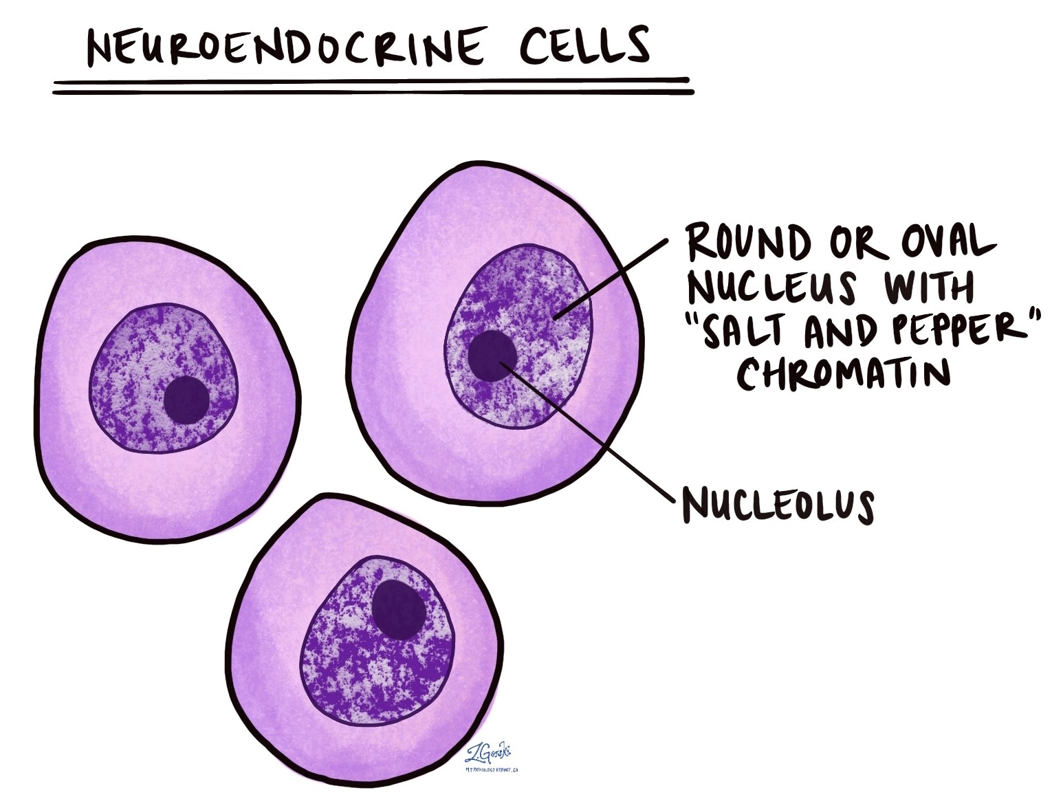
What do neuroendocrine cells look like under the microscope?
Normal neuroendocrine cells are often small and scattered, making them difficult to see on routine slides stained with hematoxylin and eosin (H&E).
When visible, these cells typically have:
-
A round nucleus (the part of the cell that contains the genetic material).
-
A distinctive pattern of chromatin (the genetic material inside the nucleus), described by pathologists as “salt and pepper”, meaning the material is evenly distributed in fine dots.
-
Occasionally, larger, darker clumps of chromatin, known as nucleoli, are visible.
This microscopic appearance is shared by both normal neuroendocrine cells and tumours made from these cells.
What are neuroendocrine tumours?
Neuroendocrine tumours are growths made up of neuroendocrine cells. These tumours can be benign (non-cancerous) or malignant (cancerous), and they can appear in many different parts of the body.
The name and behaviour of the tumour depend on:
-
Where the tumour started.
-
How the tumour looks under the microscope.
-
What hormones or proteins the tumour cells produce.
Some neuroendocrine tumours grow slowly and may not cause symptoms for years. Others grow more quickly and may cause symptoms by producing excess hormones or by spreading to other parts of the body.
Common types of neuroendocrine tumours include:
-
Carcinoid tumour – often found in the gastrointestinal tract or lungs; usually slow-growing.
-
Well-differentiated neuroendocrine tumour (NET) – a broader category that includes carcinoid tumours and other slow-growing. tumours
-
Neuroendocrine carcinoma – a more aggressive, faster-growing type of cancer made up of abnormal neuroendocrine cells.
What is neuroendocrine carcinoma?
Neuroendocrine carcinoma is a type of malignant tumour (cancer) made up of neuroendocrine cells. These cancers usually grow and spread more quickly than well-differentiated neuroendocrine tumours.
In some parts of the body, neuroendocrine carcinomas are divided into subtypes based on how the tumour cells look under the microscope:
-
Small cell neuroendocrine carcinoma – the cancer cells are small, with minimal cytoplasm and high rates of cell division. This type is commonly found in the lungs, but it can occur in other organs as well.
-
Large cell neuroendocrine carcinoma – the cancer cells are larger and more irregular in shape, but they still show neuroendocrine features.
Both types are considered high-grade cancers, meaning they grow quickly and may require aggressive treatment.
How do pathologists test for neuroendocrine cells?
To confirm that a tumour is made of neuroendocrine cells, pathologists often use a technique called immunohistochemistry. This test uses special antibodies to look for proteins made by neuroendocrine cells.
The most common proteins tested include:
-
Synaptophysin
-
Chromogranin A
-
CD56
If the tumour cells produce these proteins, it supports the diagnosis of a neuroendocrine tumour or carcinoma. Additional tests may be performed to determine the exact type of tumour and guide treatment.
Questions to ask your doctor
-
Does my tumour involve neuroendocrine cells?
-
What type of neuroendocrine tumour or carcinoma do I have?
-
Are additional tests needed to confirm the diagnosis?
-
What treatment options are available based on the type and grade of tumour?
-
Are any hormones or substances being produced by the tumour?
We are proud to partner with:
![]()