by Ashley Flaman MD and Bibianna Purgina MD FRCPC
November 10, 2023
An osteoid osteoma is a common type of non-cancerous bone tumour. These tumours are usually small (less than 2 cm) and are most frequently found in the bones of the legs, arms, spine, hands, and feet. They generally occur in children and young adults but can occasionally be found in older adults. The most common symptom is pain, which may be severe and is often worse at night.
How is this diagnosis made?
The diagnosis of osteoid osteoma can be made after a small tissue sample is removed in a procedure called a biopsy or when the entire tumour is removed in a procedure called a resection. The tissue is then sent to a pathologist for examination under the microscope. However, in some cases, the diagnosis can be made without a tissue examination because the symptoms and the appearance on X-rays are very characteristic.
This diagnosis can only be made if the tumour is less than 2 cm in size. For this reason, your pathologist may also look at your X-ray or other imaging results before making the diagnosis of osteoid osteoma. A tumour that looks similar when examined under the microscope but measures more than 2 cm is called an osteoblastoma.
What does an osteoid osteoma look like under the microscope?
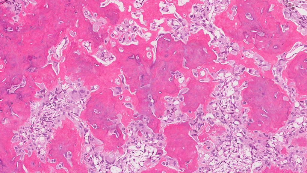
Under the microscope, an osteoid osteoma is made up of disorganized, immature new bone called osteoid surrounded by osteoblasts. Pathologists use the phrase “osteoblastic rimming” to describe osteoid surrounded by osteoblasts. This feature is important because some types of bone cancer can look similar to this tumour under the microscope, but bone cancer does not show osteoblastic rimming.

What is the typical treatment for osteoid osteoma?
Treatment options for osteoid osteoma include radio-frequency ablation or surgical removal of the tumour. A tiny piece of the tumour might also be removed by a special biopsy needle during radio-frequency ablation. That tissue will then be sent to a pathologist for examination under the microscope.
About this article
This article was written by doctors to help you read and understand your pathology report. Contact us if you have any questions about this article or your pathology report. Read this article for a more general introduction to the parts of a typical pathology report.


