by Allison Osmond, MD FRCPC
May 5, 2023
What is sebaceous carcinoma?
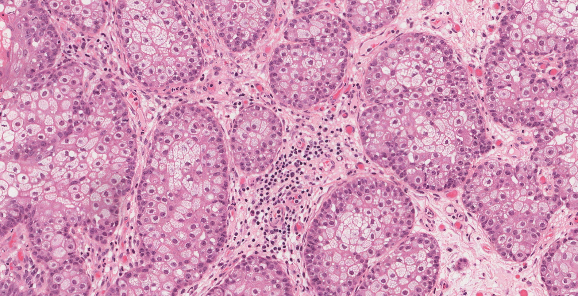
Sebaceous carcinoma is a type of skin cancer. Sebaceous carcinoma develops from specialized cells called sebocytes in the dermis and subcutaneous tissue of the skin. The cancer cells produce a fatty substance called sebum which often makes the tumour look yellow.
Where in the body is sebaceous carcinoma found?
One of the most common locations for sebaceous carcinoma is in the skin around the eye. Other locations include the face, scalp, neck, and chest.
What are the symptoms of sebaceous carcinoma?
The symptoms of sebaceous carcinoma include a painful, slow-growing, yellow lump on the skin.
What causes sebaceous cell carcinoma?
Doctors do not know exactly what causes sebaceous carcinoma. However, risk factors for developing this type of cancer include prior radiation to the skin, excessive sun exposure, and viral infections (HPV and HIV).
What genetic syndromes are associated with sebaceous cell carcinoma?
People with Muir Torre syndrome are at an increased risk of developing sebaceous carcinoma. People with this syndrome e tend to develop multiple sebaceous carcinomas and the tumours are larger than in a patient without the syndrome. People with Muir Torre syndrome also have an increased risk of developing colon cancer. If you have been diagnosed with multiple sebaceous carcinomas, your doctor may suggest a genetic test to see if you have Muir Torre syndrome.
How is sebaceous carcinoma diagnosed?
The diagnosis is usually made after a small tissue sample is removed in a procedure called a biopsy. The diagnosis can also be made after the entire tumour is removed in a procedure called an excision. If the diagnosis is made after a biopsy, your doctor will probably recommend a second surgical procedure to remove the rest of the tumour.

What is a margin and why are margins important?
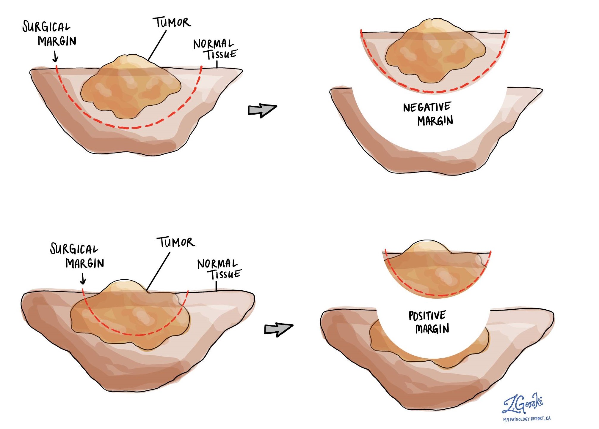
A margin is any tissue that was cut by the surgeon in order to remove the tumour from your body. Whenever possible, surgeons will try to cut tissue outside of the tumour to reduce the risk that any cancer cells will be left behind after the tumour is removed.
Your pathologist will carefully examine all the margins in your tissue sample to see how close the cancer cells are to the edge of the cut tissue. Margins will only be described in your report after the entire tumour has been removed.
A negative margin means there were no cancer cells at the very edge of the cut tissue. If all the margins are negative, most pathology reports will say how far the closest cancer cells were to a margin. The distance is usually described in millimetres.
A margin is considered positive when there are cancer cells at the very edge of the cut tissue. A positive margin is associated with a higher risk that the tumour will recur in the same site after treatment.


