by Jason Wasserman MD PhD FRCPC
January 18, 2025
HPV-associated squamous cell carcinoma of the oropharynx is a type of cancer that starts in the squamous cells lining the oropharynx, which includes parts of the throat such as the tonsils and the base of the tongue. It is linked to infection with the human papillomavirus (HPV), a common sexually transmitted virus. HPV-associated squamous cell carcinoma is different from cancers caused by other factors, like smoking or alcohol, because it has unique biological features and tends to respond better to treatment.

What are the symptoms of HPV-associated squamous cell carcinoma of the oropharynx?
The symptoms of HPV-associated squamous cell carcinoma can vary, but common ones include:
- A persistent sore throat.
- Difficulty swallowing.
- A lump in the neck (caused by swollen lymph nodes).
- Ear pain.
- Voice changes or hoarseness.
- Unexplained weight loss.
You should speak with your doctor if you experience any of these symptoms, especially for more than two weeks.
What causes HPV-associated squamous cell carcinoma of the oropharynx?
This cancer is caused by infection with high-risk types of HPV, particularly HPV-16. HPV can infect the cells in the oropharynx and cause genetic changes that lead to cancer over time. The virus spreads through direct contact, including sexual activity. It is important to note that most people who are exposed to HPV do not develop cancer. Other factors, such as smoking, may increase the risk of developing this type of cancer.
Is HPV-associated squamous cell carcinoma of the oropharynx an aggressive type of cancer?
HPV-associated squamous cell carcinoma tends to grow and spread differently than cancers not caused by HPV. While it frequently spreads to lymph nodes, it generally responds well to treatment and has a better prognosis than HPV-independent cancers. However, early detection and treatment are still important for the best outcomes.
How is this diagnosis made?
The diagnosis is made by taking a sample of tissue from the oropharynx during a procedure called a biopsy. The sample is examined under a microscope by a pathologist to look for cancerous changes. Additional tests may be performed to confirm the presence of high-risk HPV, such as immunohistochemistry for a protein called p16 or tests for HPV RNA.
What are the microscopic features of HPV-associated squamous cell carcinoma of the oropharynx?
Under the microscope, HPV-associated squamous cell carcinoma looks different from other types of squamous cell carcinoma. Unlike HPV-independent cancers, which show a gradual progression from dysplasia (abnormal surface cells) to invasive cancer, HPV-associated cancers often start in the tonsillar crypts, small folds in the tissue. The cancer cells grow as clusters or nests beneath the surface and may spread to nearby lymphoid tissue even if the surface appears unaffected.
The cancer cells typically have:
- Large nuclei (the part of the cell containing DNA) compared to the rest of the cell.
- Oval or spindle-shaped nuclei.
- Indistinct cell borders, giving the cells a “blended” appearance.
- Little or no keratin, a protein normally found in squamous cells.
These features are known as non-keratinizing squamous cell carcinoma. The surrounding tissue often contains immune cells, forming a lymphoid background.
Is HPV-associated squamous cell carcinoma of the oropharynx graded?
Unlike many other cancers, HPV-associated squamous cell carcinoma is not graded because studies have shown that grading does not predict how the cancer will behave or respond to treatment.
What is p16 and how does it aid in the diagnosis?
p16 is a protein made by cells when they are affected by high-risk types of HPV. Pathologists test for p16 using a technique called immunohistochemistry. If the cancer cells show strong staining for p16, it supports the diagnosis of HPV-associated squamous cell carcinoma.
Tests for high-risk HPV
High-risk HPV can also be identified directly using advanced laboratory tests, such as:
- In situ hybridization (ISH): This test detects HPV RNA in the cancer cells while preserving the tissue structure.
- Reverse transcription polymerase chain reaction (RT-PCR): This test detects and measures high-risk HPV RNA, proving that the virus is active in the cancer.
Tumour size
After the tumour is removed completely, it will be measured. The tumour is usually measured in three dimensions, but your report will describe only the largest dimension. For example, if the tumour measures 4.0 cm by 2.0 cm by 1.5 cm, your report will describe it as 4.0 cm. Tumour size is important for HPV-associated squamous cell carcinoma because it determines the pathologic tumour stage (pT). Larger tumours are more likely to spread to other body parts, such as lymph nodes.
Margins
Margins refer to the edges of the tissue the surgeon cut through to remove the tumour from your body. Pathologists examine these edges under a microscope to determine if any tumour cells are present. The type of margins described in your report depends on the organ involved and the type of surgery you had. Margins are only evaluated when the entire tumour has been removed.
What does it mean if a margin is negative?
A negative margin means no tumour cells were found at the edges of the removed tissue. This indicates that the tumour was entirely removed during surgery, lowering the risk of returning to the same area.
What does it mean if a margin is positive?
A positive margin means tumour cells were found right at the edge of the removed tissue. This suggests that some tumour cells may still be left behind in the body. A positive margin is associated with a higher risk of the tumour growing back in the same location after treatment. If a margin is positive, your doctor may recommend additional treatment, such as surgery, radiation, or other therapies, to help eliminate all tumour cells.
Lymph nodes
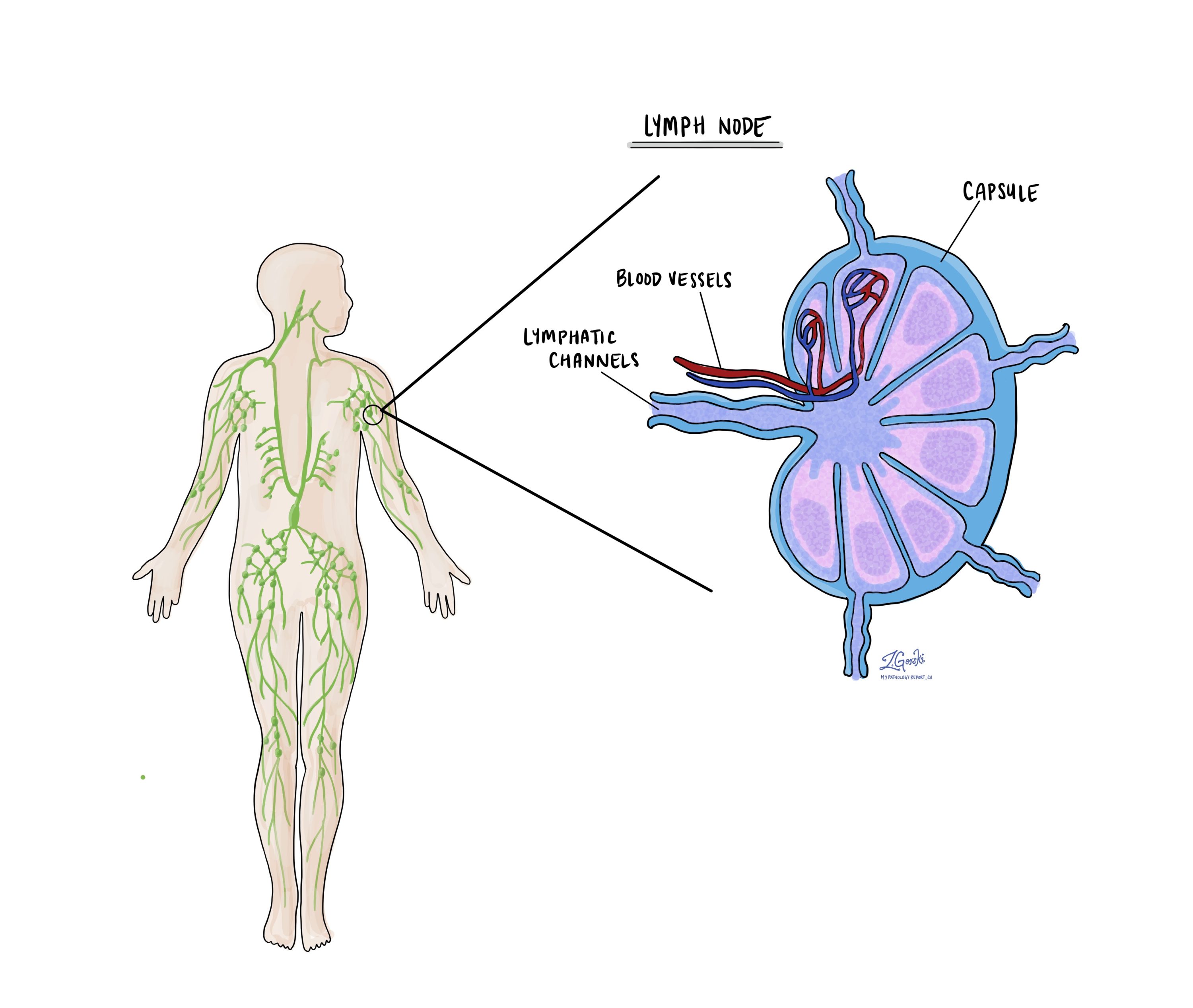
Lymph nodes are small immune organs found throughout the body. The cancer cells in HPV-associated squamous cell carcinoma frequently spread to lymph nodes in the neck. For this reason, lymph nodes are commonly removed and examined under a microscope to look for cancer cells. The movement of cancer cells from the tumour to another part of the body, such as a lymph node, is called metastasis.

Cancer cells typically spread first to lymph nodes close to the tumour, although lymph nodes far away can also be involved. For this reason, the first lymph nodes removed are usually close to the tumour. Lymph nodes further away from the tumour are typically removed only if they are enlarged, and there is a high clinical suspicion that there may be cancer cells in them.
Neck dissections
A neck dissection is a surgical procedure to remove lymph nodes from the neck. The lymph nodes removed usually come from different neck areas, and each region is called a level. The levels in the neck include 1, 2, 3, 4, and 5. Your pathology report will often describe how many lymph nodes were seen in each level sent for examination. Lymph nodes on the same side as the tumour are called ipsilateral, while those on the opposite side of the tumour are called contralateral.

How the lymph nodes will be described in your pathology report
If any lymph nodes are removed from your body, they will be examined under the microscope by a pathologist, and the examination results will be described in your report. “Positive” means that cancer cells were found in the lymph node. “Negative” indicates that no cancer cells were found. If cancer cells are found in a lymph node, the size of the largest group of cancer cells (often described as “focus” or “deposit”) may also be included in your report. Extranodal extension means that the tumour cells have broken through the capsule outside of the lymph node and spread into the surrounding tissue.
Why is the examination of lymph nodes important?
The examination of lymph nodes is important for two reasons. First, this information determines the pathologic nodal stage (pN). Second, finding cancer cells in a lymph node increases the risk that cancer cells will be found in other parts of the body in the future. As a result, your doctor will use this information when deciding if additional treatment, such as radioactive iodine, chemotherapy, radiation therapy, or immunotherapy, is required.
Pathologic stage
The pathologic stage for HPV-associated SCC of the oropharynx can only be determined after the entire tumour has been removed and sent to a pathologist for examination under the microscope. Your doctors will use the information in the pathologic stage to determine the final clinical stage.
Tumour stage (pT) for HPV-associated squamous cell carcinoma
HPV-associated SCC of the oropharynx is given a tumour stage between 1 and 4. The tumour stage is based on the size of the tumour and whether the tumour has grown to include parts of the mouth or throat outside of the oropharynx.
- T1 – The tumour is 2 cm or smaller.
- T2 – The tumour is greater than 2 cm but not larger than 4 cm.
- T3 – The tumour is larger than 4 cm but only located within the oropharynx.
- T4 – The tumour has spread into tissues outside the oropharynx, such as the deep muscles of the tongue, the larynx, or the bone of the lower jaw (the mandible).
Nodal stage (pN) for HPV-associated squamous cell carcinoma
HPV-associated SCC is given a nodal stage between 0 and 2 based on the number of lymph nodes that contain cancer cells.
- N0 – No cancer cells are found in any lymph nodes examined.
- N1 – Cancer cells are found in 1 to 4 lymph nodes examined.
- N2 – Cancer cells are found in more than 4 lymph nodes examined.
Prognosis
The prognosis for HPV-associated squamous cell carcinoma of the oropharynx is significantly better than for cancers not linked to HPV. Approximately 20% of patients experience a recurrence of the disease after treatment. However, overall survival rates are high, with 86% of patients surviving at least three years and 80% surviving at least five years after diagnosis.
Smoking can reduce the favorable prognosis, so patients are encouraged to quit smoking to improve outcomes. With proper treatment and follow-up care, many people with this diagnosis achieve excellent long-term results.


