

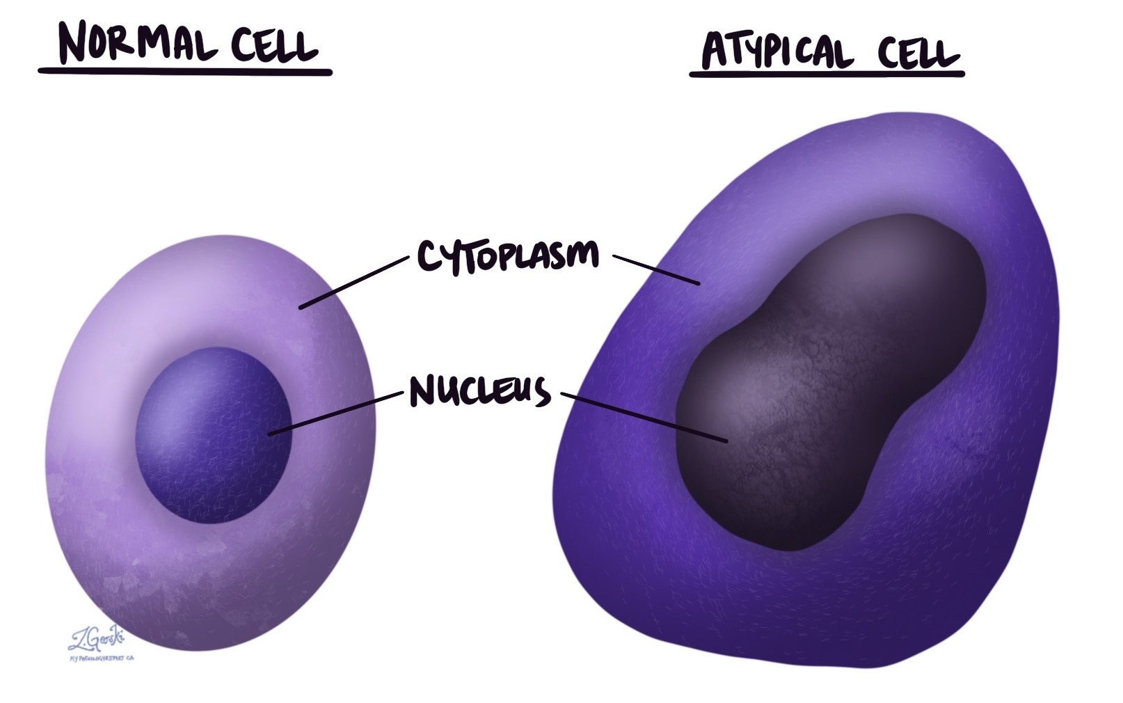
In pathology, the term atypical describes cells that look unusual or abnormal under the microscope. This term refers to changes observed in the shape, size, or structure of cells, particularly affecting the cell’s cytoplasm (the body of the cell) or the nucleus (the part that holds the genetic material).
Does atypical mean malignant?
No, atypical does not necessarily mean malignant. While malignant (cancerous) tumors often contain atypical cells, these changes can also appear in many non-cancerous conditions. Your pathologist will consider these findings along with other test results and your medical history to determine the exact meaning and significance of atypical cells in your situation.
Does atypical mean benign?
Atypical cells can sometimes be part of benign (non-cancerous) conditions. They do not automatically indicate cancer and benign conditions frequently show atypical changes. However, because atypical cells are also seen in precancerous or cancerous conditions, your doctor may recommend additional tests or follow-up to clarify their significance.
What causes cells to become atypical?
Cells may become atypical due to several reasons, including:
- Inflammation: Inflammation is your body’s natural response to injury, infection, or disease. Cells located near inflammation can appear atypical temporarily. Usually, these cells return to normal once the inflammation resolves.
- Infection: Cells infected by viruses often look atypical under the microscope. Pathologists refer to these viral changes as viral cytopathic effects. These atypical cells usually return to normal once the infection clears.
- Radiation: Radiation therapy, commonly used to treat cancer, can cause healthy cells to appear atypical. If you’ve received radiation therapy, it’s important to inform your doctor and pathologist, as these changes can persist or resolve over time.
- Precancerous diseases: Precancerous conditions, such as dysplasia or carcinoma in situ, contain atypical cells that could progress to cancer if left untreated. Recognizing these changes early helps in proper monitoring and management.
- Cancer: Cancer cells almost always appear atypical compared to healthy cells. The degree of atypical appearance helps pathologists diagnose cancer and determine its grade, influencing treatment decisions.
What do atypical cells look like under the microscope?
Under the microscope, atypical cells often show irregular shapes, sizes, or structures. They may have enlarged or unusually shaped nuclei, with changes in the appearance of the cell’s genetic material. The cell borders may appear unclear or irregular, and the cells might vary significantly from the normal surrounding cells. These microscopic features help pathologists identify atypical cells and investigate further to understand their cause and significance.
We are proud to partner with:
![]()