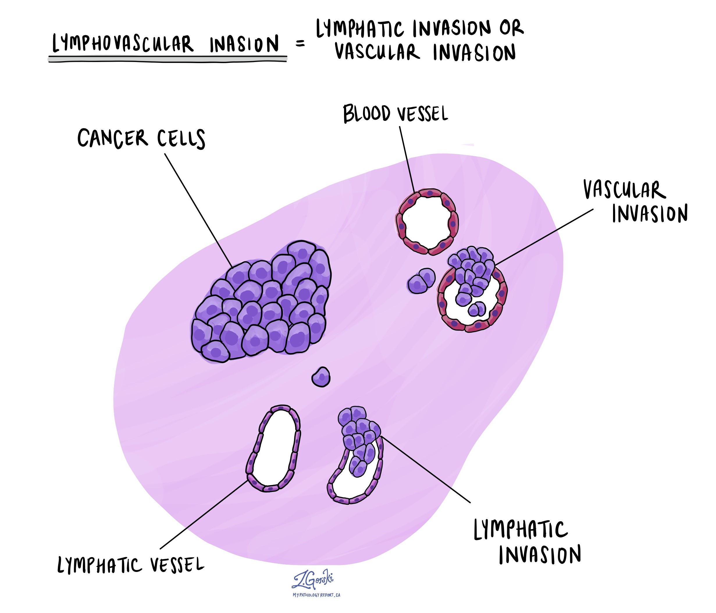
Lymphovascular invasion (LVI) means cancer cells have entered the tiny channels called lymphatic vessels or blood vessels within your body. These vessels normally carry fluid (lymph) or blood throughout your body. Once cancer cells enter these vessels, they may move away from the original tumor and reach lymph nodes or other body areas.
Does lymphovascular invasion mean the cancer has spread?
Lymphovascular invasion suggests an increased risk that cancer may spread, but it does not necessarily mean that cancer has already spread to distant parts of your body. It indicates that cancer cells have gained access to the pathways that could allow them to travel to lymph nodes or other organs. Your doctor will likely recommend further tests and careful monitoring to determine whether spreading has occurred.
What does it mean if my report says that lymphovascular invasion is positive or present?
If your pathology report says that lymphovascular invasion is “positive” or “present,” it means that cancer cells were found inside lymphatic or blood vessels when the tissue was examined under a microscope. This finding is important because it shows that your cancer has a higher potential to spread beyond its original site. Your medical team will use this information to guide surgery, chemotherapy, radiation therapy, and follow-up care decisions.
What does it mean if my report says that lymphovascular invasion is negative or not identified?
If your pathology report says that lymphovascular invasion is “negative” or “not identified,” it means that cancer cells were not seen within lymphatic or blood vessels in the tissue examined under the microscope. This finding suggests a lower likelihood that your cancer will spread beyond its original location, which may influence decisions about your treatment and follow-up care. However, your doctor will still carefully monitor your condition.
How is lymphovascular invasion different from lymphatic invasion?
Lymphatic invasion describes cancer cells entering lymphatic vessels, which are part of the lymphatic system and carry lymph fluid toward lymph nodes. In contrast, lymphovascular invasion is a broader term that includes cancer cells entering either lymphatic vessels or blood vessels. Detecting lymphovascular invasion highlights a higher risk that the cancer could spread via both the lymphatic system and the bloodstream, making it particularly important when planning treatment and monitoring.
What types of cancer typically show lymphovascular invasion?
Lymphovascular invasion can be seen in many types of cancer, including breast cancer, colorectal cancer, prostate cancer, lung cancer, and thyroid cancer, among others. Its presence is an important factor in determining the aggressiveness of the disease, potential for spreading, and appropriate treatment strategies for these cancers.
What additional tests may be performed to look for lymphovascular invasion?
Pathologists may perform additional tests to evaluate lymphovascular invasion, such as immunohistochemistry. Immunohistochemistry uses special markers to highlight lymphatic and blood vessels clearly under the microscope. For example, the marker D2-40 specifically identifies lymphatic vessels, while markers such as ERG or CD31 highlight blood vessels. These tests help confirm whether cancer cells have entered these vessels and assist in accurate staging and treatment planning.


