

Section Editor: Trevor Flood MD FRCPC
October 10, 2026
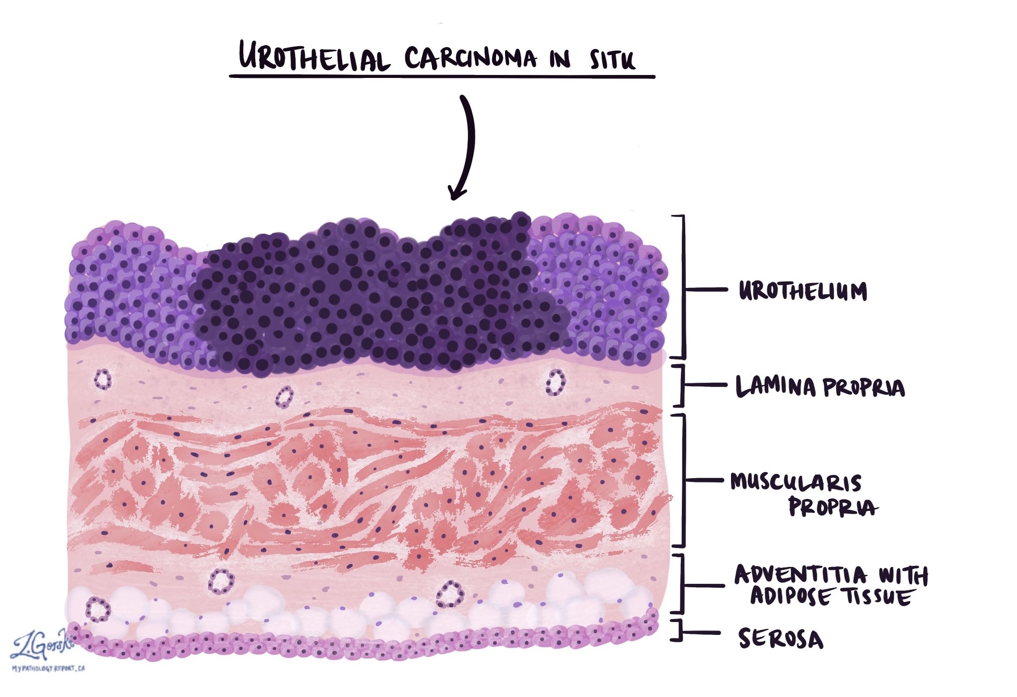
Urothelial carcinoma in situ, often shortened to CIS, is a type of cancer that starts in the urothelial cells lining the urinary tract. The term in situ means the cancer cells are still confined to the lining and have not grown into the tissue below. Unlike papillary tumors, which grow as finger-like projections, urothelial carcinoma in situ grows as a flat layer of abnormal cells.
Urothelial carcinoma in situ most often starts in the bladder, but it can also involve other parts of the urinary tract and the ducts of the prostate. It can be found on its own or alongside another bladder tumor. Although it is non-invasive, it is always high-grade and often grows into the bladder wall without treatment.
This article explains how doctors diagnose urothelial carcinoma in situ and what your pathology report says about its extent, where it was found, and its stage. It also explains why treatment is needed even though the cancer has not invaded.
Who gets urothelial carcinoma in situ?
Urothelial carcinoma in situ is mainly found in older adults, and most people are in their 60s or 70s when it is diagnosed. Like other bladder cancers, it is about four times more common in men than in women. It is most often found together with a papillary or invasive urothelial carcinoma, and less often on its own.
What causes urothelial carcinoma in situ?
Smoking is the most important cause of urothelial carcinoma in situ, because chemicals from tobacco smoke are filtered into the urine and damage the lining of the bladder. Long-term exposure to certain industrial chemicals, such as aromatic amines once used in dye and rubber manufacturing, also increases risk. Other causes include arsenic in drinking water and earlier treatment with radiation to the pelvis or with the chemotherapy drug cyclophosphamide. Stopping smoking lowers the chance of new tumors forming after treatment.
What are the symptoms of urothelial carcinoma in situ?
Urothelial carcinoma in situ often causes symptoms that feel like a bladder infection. These include burning during urination and needing to urinate often or urgently, even when urine tests show no infection. Some people have blood in the urine, which may be visible or found only on a urine test. Others have no symptoms, and the cancer is found during tests for another bladder tumor.
How is the diagnosis made?
The diagnosis of urothelial carcinoma in situ usually begins with urine tests and a look inside the bladder. In urine cytology, a pathologist looks for cancer cells in a urine sample. Because urothelial carcinoma in situ cells are loosely attached and highly abnormal, they often shed into the urine and can be detected by this test. Our article on cytology reports explains this type of test.
A urologist then looks inside the bladder with a thin camera, a procedure called cystoscopy. Urothelial carcinoma in situ may look like a flat, red, velvety patch, or the lining may look normal. Some centers use a special blue light during cystoscopy, which makes these flat areas easier to see. The urologist takes small samples, called biopsies, from any abnormal areas. The urologist may also take samples from normal-looking areas to check how widely the cancer has spread through the lining.
Under the microscope, a pathologist sees urothelial cells that are large, dark, and very different from normal cells. Many of the cells are dividing. Because the cells stick together poorly, they often fall away from the lining. In that case, your report may say the lining is “denuded,” meaning only a few cancer cells remain on the surface.
Urothelial carcinoma in situ can be hard to distinguish from cells that look abnormal because of inflammation or earlier treatment, called reactive changes. In these cases, the pathologist may perform immunohistochemistry, a test that uses stains to highlight proteins in the cells. Common stains include cytokeratin 20, p53, and CD44. The stains chosen and how laboratories report them vary, and no single stain proves the diagnosis.
Histologic grade
Urothelial carcinoma in situ is not given a separate grade, because it is high grade by definition. The cells look very different from normal urothelial cells and share the same features as high-grade invasive urothelial carcinoma. This is why urothelial carcinoma in situ is treated more seriously than a low-grade papillary tumor, even though it has not invaded.
Some reports also describe a less abnormal flat change called urothelial dysplasia. In dysplasia, the cells look abnormal but not abnormal enough to be called carcinoma in situ. It is not cancer, but it is a sign that the lining is at risk, and it is usually followed with cystoscopy.
Extent and location of urothelial carcinoma in situ
Your report for urothelial carcinoma in situ may describe whether it was found alone or with another tumor. It may also describe how much of the lining is involved and which parts of the urinary tract are affected. These findings help your team decide on treatment.
- Primary carcinoma in situ. The carcinoma in situ was found alone, without a papillary or invasive tumor.
- Carcinoma in situ with another tumor. The carcinoma in situ was found together with a papillary urothelial carcinoma or an invasive urothelial carcinoma. Its presence increases the risk that the other tumor will recur or grow deeper.
- Focal or extensive. Focal means the carcinoma in situ was found in one small area. Extensive or multifocal means it was found in several samples or over a large area of the lining, which is linked to a higher risk.
Urothelial carcinoma in situ can spread along the lining into nearby parts of the urinary tract. In men, it can grow into the prostatic urethra, which is the part of the urethra that passes through the prostate. It can also extend into the small ducts of the prostate. Your report will indicate whether these areas are involved, because this can change treatment and staging.
Urothelial carcinoma in situ can also involve the ureters or the lining of the kidney. For this reason, imaging and sometimes urine samples from each ureter are used to check the upper urinary tract.
Margins
A biopsy or TURBT removes urothelial carcinoma in situ in small pieces, so margins are not reported on these samples. Margins become important if the bladder is removed in an operation called a cystectomy. The pathologist then checks the cut ends of the ureters and the urethra, because carcinoma in situ can spread along the lining to these areas.
- Negative margin. No cancer cells, including carcinoma in situ, are seen at the cut ends.
- Positive margin for carcinoma in situ. Carcinoma in situ is seen at the cut end of a ureter or the urethra. This is linked to a higher chance of the cancer coming back in the remaining lining, which is checked during follow-up.
Our article on the radical cystectomy pathology report describes this operation.
Pathologic stage (pTNM)
We assign the pathologic stage for urothelial carcinoma in situ using the TNM system from the AJCC Cancer Staging Manual, 8th edition. Because the cancer cells have not grown into the underlying tissue, urothelial carcinoma in situ cannot spread to lymph nodes or distant organs on its own.
Tumor stage (pT)
- pTis. Urothelial carcinoma in situ is present in the bladder, and no invasive cancer was found.
- Staged by the invasive tumor. If carcinoma in situ is found with an invasive urothelial carcinoma, the stage is based on how deeply the invasive tumor has grown. Your report may still mention the carcinoma in situ separately.
- pTis pu and pTis pd. These stages describe carcinoma in situ in the prostatic urethra (pu) or in the prostatic ducts (pd). They come from the AJCC staging rules for the urethra.
Nodal stage (pN)
Lymph nodes are not removed during a biopsy or TURBT, so reports for urothelial carcinoma in situ usually do not include a nodal stage. If lymph nodes are removed during a cystectomy, the nodal stage is described in our article on invasive urothelial carcinoma.
What is the prognosis?
The prognosis for urothelial carcinoma in situ depends mostly on whether it responds to treatment. Because the cancer has not invaded, most people do well. However, without treatment, it often grows into the bladder wall.
- Survival. About 98 in 100 people with non-invasive bladder cancer, including carcinoma in situ, are alive five years after diagnosis, and about 2 in 100 are not.
- Without treatment. Before BCG was available, about half of people with carcinoma in situ developed cancer in the muscle layer of the bladder.
- Response to BCG. About 7 in 10 people have no detectable cancer after a first course of BCG, but in some, the cancer later comes back.
- Long-term risk. In one large group treated with BCG, about 7 in 10 had no cancer growing deeper ten years later, and about 3 in 10 did.
These numbers are averages for large groups of people and don’t tell you what will happen to you. Features in your report that are linked to a higher chance of the cancer coming back or growing deeper include:
- Extensive or multifocal disease. Carcinoma in situ found in many samples or over a large area.
- Involvement of the prostatic urethra or ducts. These areas are harder for bladder medication to reach.
- A papillary or invasive tumor alongside it. Especially a high-grade pT1 tumor.
- Cancer that persists or returns after BCG. This is the strongest sign that you need other treatment.
What happens after the diagnosis?
After a diagnosis of urothelial carcinoma in situ, your care team uses the extent, location, and any other tumor in your report to plan treatment. Because it is high-grade, urothelial carcinoma in situ places bladder cancer in the high-risk group. Treatment aims to destroy the cancer cells in the lining and prevent invasion.
- BCG. BCG is a weakened bacterium that stimulates the immune system. It is placed in the bladder through a catheter, usually weekly for six weeks, followed by maintenance treatments for up to three years.
- Durvalumab with BCG. In May 2026, the FDA approved durvalumab, an immunotherapy drug, to be given with BCG to some people with high-risk disease who have not had BCG before.
- Checking the response. Cystoscopy, urine cytology, and often biopsies are done about three months after BCG starts to see whether the cancer has gone.
- Removing the bladder. If the cancer persists or returns after BCG, removing the bladder, called a cystectomy, is often discussed. It may also be considered first for some people with very extensive disease.
Several FDA-approved medicines treat carcinoma in situ that has not responded to BCG. They are used for people who cannot have, or choose not to have, a cystectomy. These include:
- Pembrolizumab. An immunotherapy drug given intravenously, approved in January 2020.
- Nadofaragene firadenovec. A gene therapy placed in the bladder, approved in December 2022.
- Nogapendekin alfa inbakicept. An immune-stimulating drug given with BCG, approved in April 2024.
- Gemcitabine intravesical system. A small device that slowly releases chemotherapy inside the bladder, approved in September 2025.
In the studies that led to these approvals, between about 4 and 8 in 10 people had no detectable cancer after treatment, but in many it later came back. Approval and access to these drugs differ by country, and your care team can explain what applies where you live. Follow-up includes cystoscopy and urine cytology every few months at first, and regular checks of the upper urinary tract.
Questions to ask your doctor
- Was the carcinoma in situ found on its own or with another tumor?
- Is it focal, or is it extensive or multifocal?
- Does it involve my prostatic urethra or prostate ducts?
- Was any invasive cancer found in the samples?
- Were special stains used to confirm the diagnosis?
- What is the stage of my cancer?
- Will I receive BCG, and how long will the treatment last?
- Am I a candidate for durvalumab with BCG, or for a clinical trial?
- How will we know whether BCG has worked?
- What are my options if the cancer does not respond to BCG?
- Should my ureters and kidneys be checked for carcinoma in situ?
- How often will I need cystoscopy and urine cytology?
Related articles on MyPathologyReport.com
We are proud to partner with:


🔍 Search MyPathologyReport
Type what you see on your report — for example a diagnosis or test name