

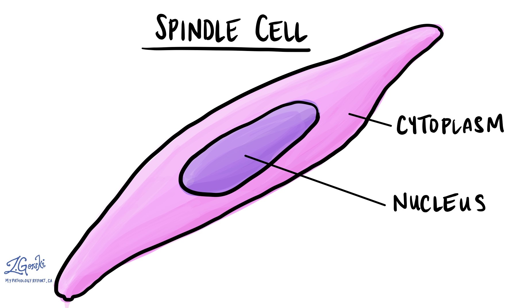
A malignant spindle cell neoplasm is a type of cancer made up of spindle-shaped cells. Under the microscope, these cells look long and narrow, similar to the shape of a spindle (a pointed rod used for spinning thread).
This diagnosis describes how the cells look under the microscope but does not identify the exact type of cancer. Many different kinds of tumours can have spindle-shaped cells, so additional testing is almost always needed to determine the precise diagnosis.
Is it a type of cancer?
Yes. The word malignant means that the tumour is cancerous, which means it can invade nearby tissues and may spread (metastasize) to other parts of the body.
However, the specific behaviour and aggressiveness of the cancer depend on its exact type. Some malignant spindle cell tumours grow slowly, while others behave more aggressively. Identifying the specific type of tumour helps doctors choose the most effective treatment and estimate the likely outcome (prognosis).
When is this diagnosis made?
This diagnosis is usually made after a biopsy, a procedure in which a small piece of tissue is removed from the tumour and examined under a microscope.
Pathologists use the term malignant spindle cell neoplasm when the tumour clearly looks cancerous but there is not enough information from the biopsy to determine its exact type. This can happen when:
-
The biopsy sample is small or contains damaged tissue.
-
The tumour cells look very abnormal, making it hard to tell what type of tissue they came from.
-
Special tests are still pending.
Features that suggest malignancy include:
-
Increased mitotic activity (cell division).
-
Areas of necrosis (dead tumour tissue).
-
Invasion into nearby tissue or blood vessels.
When more tissue becomes available — for example, after the entire tumour is surgically removed — a more specific diagnosis can often be made.
What types of tumours can look like a malignant spindle cell neoplasm?
Several types of cancers can appear spindle-shaped under the microscope.
Common examples include:
-
Sarcomas: These cancers start in connective tissues such as bone, muscle, or fat. Common types of sarcomas include leiomyosarcoma, angiosarcoma, and liposarcoma.
-
Spindle cell squamous cell carcinoma: A type of squamous cell carcinoma that develops a spindle-shaped appearance. It is often seen in organs such as the larynx, lung, or skin.
-
Malignant peripheral nerve sheath tumour (MPNST): A rare cancer that begins in the protective lining of nerves. It may occur on its own or in people with neurofibromatosis type 1 (NF1).
-
Gastrointestinal stromal tumour (GIST): A cancer that develops in the wall of the digestive tract, most often in the stomach or small intestine.
Each of these tumour types has its own pattern of growth, typical locations, and genetic features.
How do pathologists determine the exact type of tumour?
Pathologists use a combination of tests and observations to identify the specific type of malignant spindle cell tumour. These may include:
-
Microscopic features: The overall appearance, pattern, and location of the tumour.
-
Immunohistochemistry (IHC): A test that uses special stains to look for proteins (markers) inside the tumour cells. Different types of tumours produce different proteins.
-
Molecular or genetic testing: Tests such as FISH, PCR, or next-generation sequencing (NGS) can identify characteristic genetic mutations or rearrangements.
-
Clinical information: The tumour’s location, size, and behaviour in the body help narrow down the possibilities.
Combining these results allows the pathologist to make a final diagnosis, which is essential for planning treatment.
What is the prognosis for someone diagnosed with a malignant spindle cell neoplasm?
The prognosis (expected outcome) depends on several factors, including:
-
The specific type of tumour identified after testing.
-
The grade of the tumour (how aggressive it looks under the microscope).
-
The tumour size and whether it has spread to nearby tissues or other organs.
-
The location of the tumour in the body.
In general, early detection and complete removal of the tumour improve the chances of successful treatment.
Questions to ask your doctor
-
What type of malignant spindle cell neoplasm do I have?
-
Has additional testing been ordered to confirm the diagnosis?
-
What did the immunohistochemistry or molecular test results show?
-
Has the tumour spread to nearby tissues or other parts of the body?
-
What are my treatment options?
-
What is the prognosis for this type of tumour?
-
Will I need further biopsies or imaging studies?
We are proud to partner with:
![]()