by Jason Wasserman MD PhD FRCPC
January 23, 2024
Background:
Alveolar soft part sarcoma is a rare type of cancer that typically starts in the body’s soft tissues, including muscles, fat, and connective tissues. Despite its name, alveolar soft part sarcoma does not start in the lung’s alveolar structures, as one might think from the word “alveolar.” Instead, it gets its name from the way the tumour cells are arranged in small nests that resemble the small air sacs in the lungs.
What are the symptoms of alveolar soft part sarcoma?
The symptoms of alveolar soft part sarcoma can vary depending on where the tumour is located. Commonly, it manifests as a painless, slow-growing lump or mass in the affected area. It most frequently occurs in the legs or arms but can also be found in the head, neck, chest, or abdomen. Symptoms might only become noticeable as the tumour grows and affects surrounding tissues or organs.
What causes alveolar soft part sarcoma?
The exact cause of alveolar soft part sarcoma is not well understood. Like many cancers, it’s thought to result from genetic changes in the body’s cells that lead to uncontrolled growth. However, it’s important to note that these changes are usually not inherited but occur randomly.
What genetic changes are found in alveolar soft part sarcoma?
Alveolar soft part sarcoma is associated with a specific genetic change known as a chromosomal rearrangement. This involves the fusion of part of chromosome X (Xp11) with chromosome 17 (17q25), resulting in the ASPSCR1-TFE3 gene fusion. This fusion is believed to play a key role in the development of the tumour.
How is this diagnosis made?
Diagnosing alveolar soft part sarcoma typically involves a combination of medical imaging and a biopsy. Imaging tests like MRI or CT scans can detect the presence of a mass and its size and location. A biopsy, where a small tumour sample is removed and examined under a microscope, is important for confirming the diagnosis.
Your pathology report for alveolar soft part sarcoma:
Microscopic features of this tumour
Under the microscope, alveolar soft part sarcoma cells have a distinctive appearance. They are often arranged in nests or clusters separated by thin walls of blood vessels, resembling the alveoli in the lungs. The cells are usually large, and the cytoplasm (body) of the cell can appear clear, eosinophilic (pink), or granular. The nucleus (the part of the cell that holds the genetic material) is typically round and prominent clumps of genetic material called nucleoli are often seen. Mitotic figures (dividing cells) may be seen, but the mitotic rate is usually low (the number of mitotic figures in a given tissue area).

Immunohistochemistry
Immunohistochemistry (IHC) involves using special stains (antibodies) that react with specific proteins in the tumour cells. In the case of alveolar soft part sarcoma, these tests can highlight certain proteins that are typically found in the tumour cells, helping to distinguish alveolar soft part sarcoma from other types of tumours that can look similar when examined under the microscope.
- TFE3 protein: Alveolar soft part sarcoma cells usually show strong expression of the TFE3 protein. TFE3 immunostaining is particularly useful as it is associated with the TFE3 gene rearrangement characteristic of alveolar soft part sarcoma.
Molecular tests
Molecular tests on alveolar soft part sarcoma tissue focus on identifying the specific chromosomal changes that are characteristic of this cancer. The most significant change is a translocation between chromosomes X and 17, which creates the ASPSCR1-TFE3 gene fusion. The details of these tests include:
- Fluorescence in situ hybridization (FISH): This test uses fluorescent DNA that specifically binds to the ASPSCR1-TFE3 gene fusion on the chromosomes in the tumour cells. The presence of this fusion is a strong indicator of alveolar soft part sarcoma.
- Next-generation sequencing (NGS): NGS is a powerful molecular test that can simultaneously look for thousands of genetic changes in a small sample of tissue. It can detect the genetic fusion of ASPSCR1 and TFE3. This test is highly sensitive and specific for the genetic changes seen in alveolar soft part sarcoma.
- Reverse transcriptase polymerase chain reaction (RT-PCR): RT-PCR can detect the specific genetic fusion of ASPSCR1 and TFE3 at the molecular level. This test is highly sensitive and specific for the genetic changes seen in alveolar soft part sarcoma.
Tumour grade
All alveolar soft part sarcomas are considered high grade tumours. This means they tend to grow and spread more aggressively than low grade tumours.
Treatment effect
If you received chemotherapy and/or radiation therapy before the operation to remove the tumour, your pathologist will examine all the tissue sent to pathology to see how much of the tumour was still alive at the time it was removed from the body. Pathologists use the term viable to describe tissue that was still alive when it was removed from the body. In contrast, pathologists use the term non-viable to describe tissue that was not alive when it was removed from the body. Most commonly, your pathologist will describe the percentage of tumours that is non-viable.
Perineural invasion
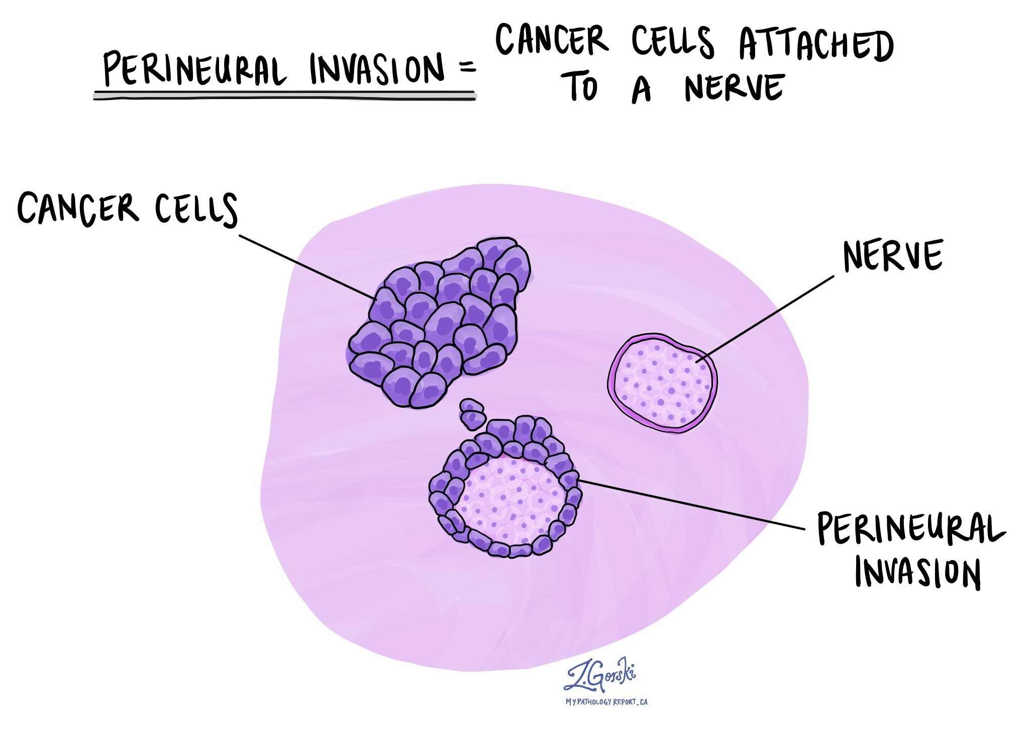
Pathologists use the term “perineural invasion” to describe a situation where cancer cells attach to or invade a nerve. “Intraneural invasion” is a related term that specifically refers to cancer cells found inside a nerve. Nerves, resembling long wires, consist of groups of cells known as neurons. These nerves, present throughout the body, transmit information such as temperature, pressure, and pain between the body and the brain. The presence of perineural invasion is important because it allows cancer cells to travel along the nerve into nearby organs and tissues, raising the risk of the tumour recurring after surgery.

Lymphovascular invasion
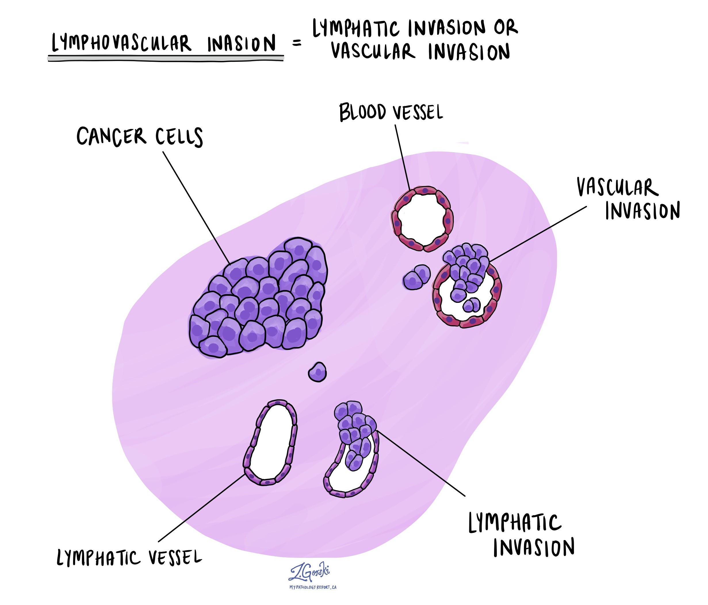
Lymphovascular invasion occurs when cancer cells invade a blood vessel or lymphatic channel. Blood vessels, thin tubes that carry blood throughout the body, contrast with lymphatic channels, which carry a fluid called lymph instead of blood. These lymphatic channels connect to small immune organs known as lymph nodes scattered throughout the body. Lymphovascular invasion is important because it enables cancer cells to spread to other body parts, including lymph nodes or the lungs, via the blood or lymphatic vessels. As such, lymphovascular invasion is associated with an increased risk of developing metastatic disease.

Margins
In pathology, a margin is the edge of tissue removed during tumour surgery. The margin status in a pathology report is important as it indicates whether the entire tumour was removed or if some was left behind. This information helps determine the need for further treatment.
Pathologists typically assess margins following a surgical procedure, like an excision or resection, that removes the entire tumour. Margins aren’t usually evaluated after a biopsy, which removes only part of the tumour. The number of margins reported and their size—how much normal tissue is between the tumour and the cut edge—vary based on the tissue type and tumour location.
Pathologists examine margins to check if tumour cells are present at the tissue’s cut edge. A positive margin, where tumour cells are found, suggests that some cancer may remain in the body. In contrast, a negative margin, with no tumour cells at the edge, suggests the tumour was fully removed. Some reports also measure the distance between the nearest tumour cells and the margin, even if all margins are negative.

Lymph nodes
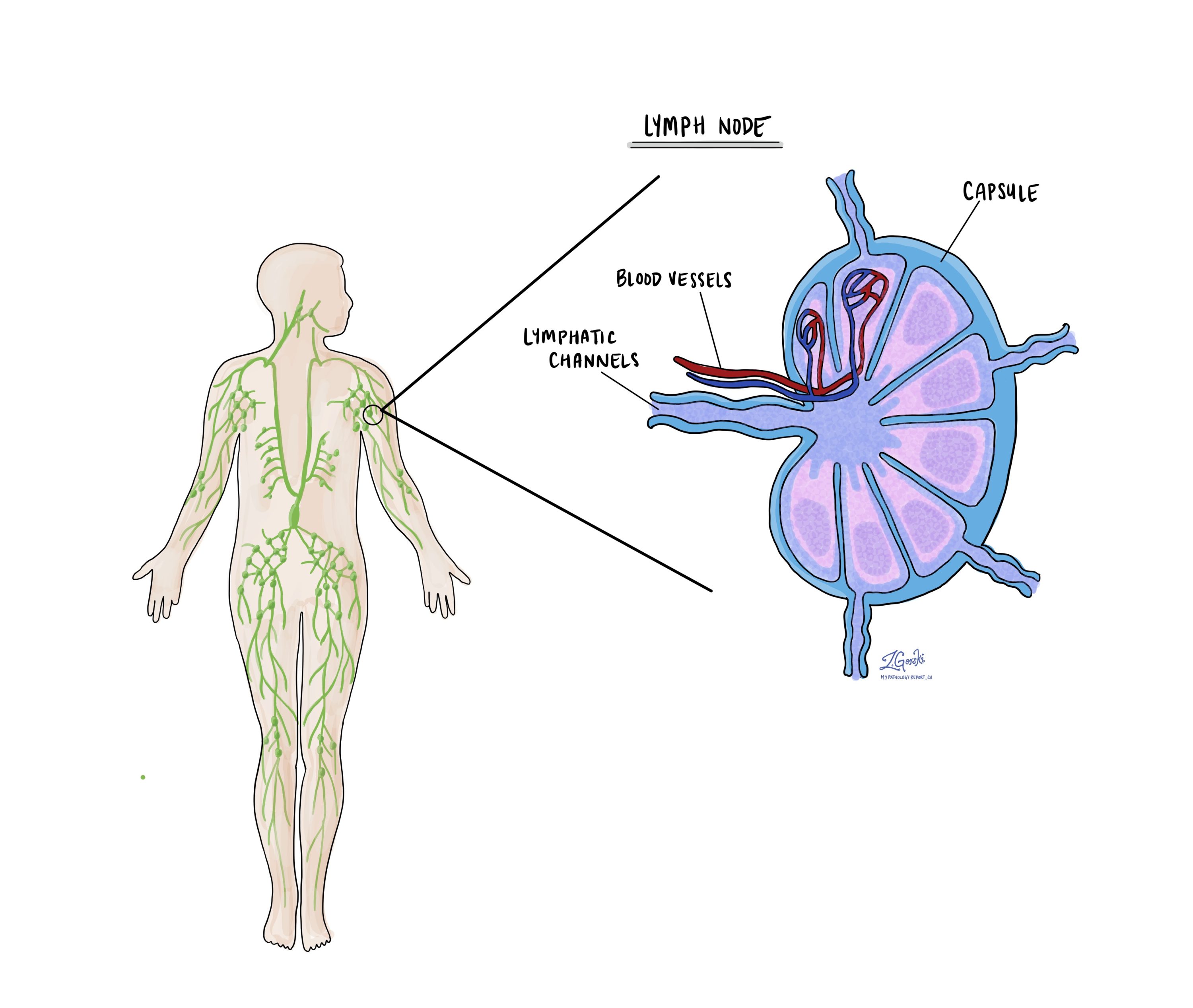
Lymph nodes are small immune organs found throughout the body. Cancer cells can spread from a tumour to lymph nodes through small lymphatic vessels. For this reason, lymph nodes are commonly removed and examined under a microscope to look for cancer cells. The movement of cancer cells from the tumour to another part of the body, such as a lymph node, is called a metastasis.

Cancer cells typically spread first to lymph nodes close to the tumour, although lymph nodes far away from the tumour can also be involved. For this reason, the first lymph nodes removed are usually close to the tumour. Lymph nodes further away from the tumour are only typically removed if they are enlarged and there is a high clinical suspicion that there may be cancer cells in the lymph node.
If any lymph nodes were removed from your body, they will be examined under the microscope by a pathologist, and the results of this examination will be described in your report. The examination of lymph nodes is important for two reasons. First, this information determines the pathologic nodal stage (pN). Second, finding cancer cells in a lymph node increases the risk that cancer cells will be found in other parts of the body in the future. As a result, your doctor will use this information when deciding if additional treatment, such as chemotherapy, radiation therapy, or immunotherapy, is required.
Some helpful definitions:
- Positive: Positive means that cancer cells were found in the lymph node being examined.
- Negative: Negative means that no cancer cells were found in the lymph node being examined.
- Deposit: The term deposit describes a group of cancer cells inside a lymph node. Some reports include the size of the largest deposit. A similar term is “focus”.
- Extranodal extension: Extranodal extension means that the tumour cells have broken through the capsule on the outside of the lymph node and have spread into the surrounding tissue.

Pathologic stage (pTNM)
The pathologic stage for alveolar soft part sarcoma is based on the TNM staging system, an internationally recognized system created by the American Joint Committee on Cancer. This system uses information about the primary tumour (T), lymph nodes (N), and distant metastatic disease (M) to determine the complete pathologic stage (pTNM). Your pathologist will examine the tissue submitted and give each part a number. In general, a higher number means a more advanced disease and a worse prognosis.
Tumour stage (pT)
The tumour stage for alveolar soft part sarcoma varies based on the body part involved. For example, a 5-centimetre tumour that starts in the head will be given a different tumour stage than a tumour that starts deep in the back of the abdomen (the retroperitoneum). However, in most body sites, the tumour stage includes the tumour size and whether the tumour has grown into surrounding body parts.
Head and neck
- T1 – The tumour is no greater than 2 centimetres in size.
- T2 – The tumour is between 2 and 4 centimetres in size.
- T3 – The tumour is greater than 4 centimetres in size.
- T4 – The tumour has grown into surrounding tissues such as the bones of the face or skull, the eye, the larger blood vessels in the neck, or the brain.
Chest, back, or stomach and the arms or legs (trunk and extremities)
- T1 – The tumour is no greater than 5 centimetres in size.
- T2 – The tumour is between 5 and 10 centimetres in size.
- T3 – The tumour is between 10 and 15 centimetres in size.
- T4 – The tumour is greater than 15 centimetres in size.
Abdomen and organs inside the chest (thoracic visceral organs)
- T1 – The tumour is only seen in one organ.
- T2 – The tumour has grown into the connective tissue surrounding the organ from which it started.
- T3 – The tumour has grown into at least one other organ.
- T4 – Multiple tumours are found.
Retroperitoneum (the space at the very back of the abdominal cavity)
- T1 – The tumour is no greater than 5 centimetres in size.
- T2 – The tumour is between 5 and 10 centimetres in size.
- T3 – The tumour is between 10 and 15 centimetres in size.
- T4 – The tumour is greater than 15 centimetres in size.
Tissue around the eye (orbit)
- T1 – The tumour is no greater than 2 centimetres in size.
- T2 – The tumour is greater than 2 centimetres in size but has not grown into the bones surrounding the eye.
- T3 – The tumour has grown into the bones surrounding the eye or other bones of the skull.
- T4 – The tumour has grown into the eye (the globe) or the surrounding tissues such as the eyelids, sinuses, or brain.
Nodal stage (pN)
Alveolar soft part sarcoma is given a nodal stage of 0 or 1 based on the presence or absence of tumour cells in one or more lymph nodes. If no tumour cells are seen in any lymph nodes, the nodal stage is N0. If no lymph nodes are sent for pathological examination, the nodal stage cannot be determined, and the nodal stage is listed as NX. If tumour cells are found in any lymph nodes, then the nodal stage is listed as N1.
About this article
Doctors wrote this article to help you read and understand your pathology report. If you have additional questions, contact us.

