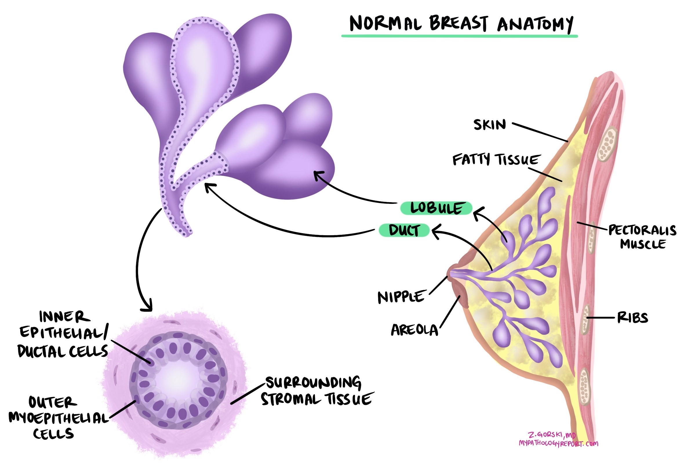
Invasive ductal carcinoma is the most common type of breast cancer. It originates from epithelial cells lining the ducts of the breast and spreads into the surrounding breast tissue. If not treated, invasive ductal carcinoma can spread to other body parts, such as the lymph nodes, bones, and lungs. Another name for this type of cancer is invasive breast carcinoma.
What are the symptoms of invasive ductal carcinoma?
The most common symptom of invasive ductal carcinoma is a new lump or mass in the breast. These lumps are often hard and irregular in shape but can also be soft or round. Some people notice changes in the size or shape of the breast, dimpling or redness of the skin, or changes in the nipple, such as inversion or discharge, especially if it is bloody. Although breast pain is usually caused by non-cancerous conditions, some patients may experience persistent pain in one area of the breast. Swelling of the breast, even without a lump, and enlarged lymph nodes under the arm or near the collarbone may also be signs of invasive ductal carcinoma.
What causes invasive ductal carcinoma?
How is this diagnosis made?
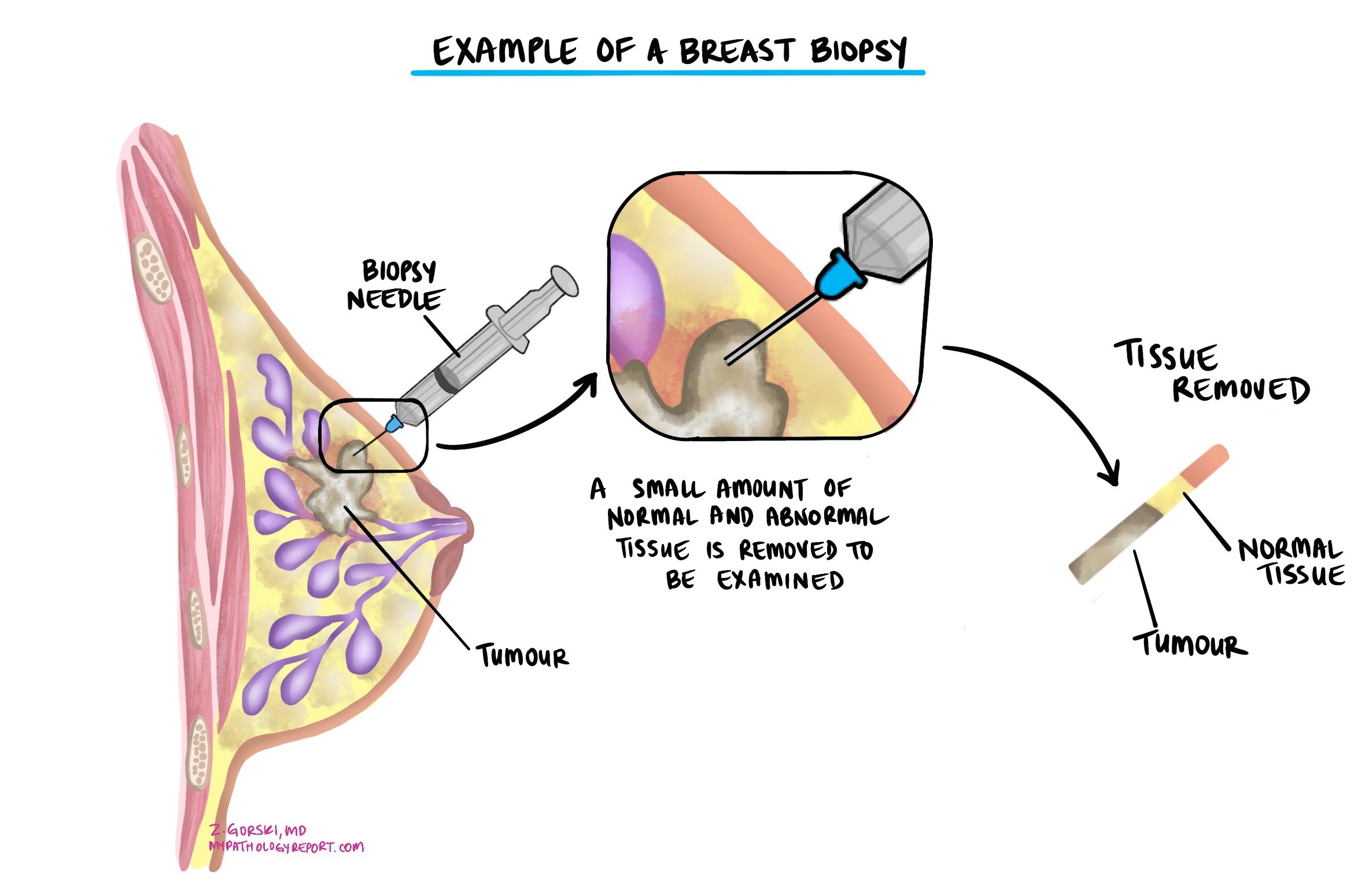
The diagnosis of invasive ductal carcinoma is usually made after a small sample of the tumour is removed in a procedure called a biopsy. The tissue is then sent to a pathologist for examination under a microscope. You may then be offered additional surgery to remove the tumour altogether.
Nottingham histologic grade
The Nottingham histologic grade, also known as the modified Scarff-Bloom-Richardson grade, is a system used by pathologists to evaluate breast cancer under the microscope. It helps determine the aggressiveness of the tumour and provides important information for planning treatment. The grade is based on how different the cancer cells look from normal breast cells and how quickly they are growing.
To calculate the grade, pathologists examine three features of the cancer:
- Tubule formation: This measures the extent to which cancer cells form structures resembling normal breast glands. If most of the cells form tubules, the tumour gets a lower score. Fewer tubules mean a higher score.
- Nuclear pleomorphism: This describes the variation in the appearance of cancer cells’ nuclei (the part of the cell that contains DNA) compared to normal cells. The score is low if the nuclei are uniform and similar to those of normal cells. If they are very different and irregular, the score is higher.
- Mitotic count: This measures the number of cancer cells that are actively dividing. Cells that are dividing undergo a process called mitosis and are referred to as mitotic figures. A higher number of dividing cells indicates that the tumour is growing quickly, resulting in a higher score.
Each feature is scored from 1 to 3, with 1 indicating a level close to normal and 3 indicating a more abnormal level. The scores are added together to give a total score between 3 and 9, which determines the grade.

The total score places the tumour into one of three grades:
- Grade 1 (Low grade): Total score of 3 to 5. Cancer cells often resemble normal cells and typically grow at a slow rate.
- Grade 2 (Intermediate grade): Total score of 6 to 7. The cancer cells show more differences from normal and grow at a moderate rate.
- Grade 3 (High grade): Total score of 8 to 9. Cancer cells appear distinctly different from normal cells and tend to grow more rapidly.
The grade helps doctors predict how aggressive the cancer will likely be. Grade 1 cancers often grow slowly and may have a better outcome. Grade 3 cancers can grow and spread more quickly and may require more aggressive treatment. Your doctor will use the grade and other factors, such as tumour size and whether cancer is found in lymph nodes, to guide treatment decisions.
Breast cancer biomarkers
Estrogen receptor (ER) and progesterone receptor (PR)
Hormone receptors are proteins found in some breast cancer cells. The two main types tested are estrogen receptor (ER) and progesterone receptor (PR). Cancer cells with these receptors utilize hormones such as estrogen and progesterone to promote growth and division. Testing for ER and PR helps guide treatment and predict prognosis.
Your pathology report will typically include:
-
Percentage of positive cells: For example, “80% ER-positive” means 80% of cancer cells have estrogen receptors.
-
Intensity of staining: Reported as weak, moderate, or strong, this indicates the number of receptors present in the cancer cells.
-
Overall score (Allred or H-score): This combines percentage and intensity, with higher scores indicating a better response to hormone therapy.
Cancer cells are described as hormone receptor-positive if ER or PR is present in at least 1% of cells. These cancers often grow more slowly, are less aggressive, and typically respond well to hormone-blocking therapies, such as tamoxifen or aromatase inhibitors (e.g., anastrozole, letrozole, or exemestane). Hormone therapy helps reduce the chance of cancer recurrence.
Tumours with ER positivity between 1% and 10% are considered ER low positive. These cancers still usually respond better to hormone therapy compared to ER-negative cancers.
HER2 testing by immunohistochemistry
Immunohistochemistry (IHC) is a test pathologists use to measure the amount of HER2 protein on the surface of breast cancer cells. HER2 (human epidermal growth factor receptor 2) is a protein that helps control cell growth. Some breast cancers make too much HER2 protein, which can cause the cells to grow and divide more quickly.
To perform the test, the pathologist applies special antibodies that bind to HER2 proteins on a small sample of tumor tissue. A coloured dye makes these antibodies visible under the microscope. By looking at the intensity (strength) and pattern of the staining, the pathologist assigns a score that reflects how much HER2 protein is present on the cancer cells.
HER2 immunohistochemistry scores
Your pathology report will describe the HER2 IHC result as a score from 0 to 3+, with additional designations for HER2-low and HER2-ultralow expression:
- 0 (negative) – No visible membrane staining, meaning no detectable HER2 protein. Tumors with this pattern are HER2-negative. Targeted HER2 therapies are not effective for these cancers.
- 0+ (ultralow) – Faint or barely visible incomplete membrane staining in 10% or fewer of the tumor cells. This pattern is called HER2-ultralow. Although still considered HER2-negative, some studies suggest these tumors may respond to new targeted therapies such as trastuzumab-deruxtecan in the metastatic (advanced) setting. Tumors with no staining at all remain ineligible for this treatment.
- 1+ (low) – Faint, incomplete membrane staining in more than 10% of the cancer cells. These tumors are HER2-low and are considered HER2-negative by traditional criteria. However, in the metastatic setting, HER2-low cancers may also be eligible for therapies that target low levels of HER2 protein.
- 2+ (equivocal) – Weak to moderate complete membrane staining in more than 10% of the tumor cells. This result is considered borderline, and further testing—usually in situ hybridization (ISH)—is needed to determine if the cancer is truly HER2-positive or HER2-negative.
If the ISH test is negative (showing no HER2 gene amplification), the result is categorized as HER2-low.
- 3+ (positive) – Strong, complete membrane staining in more than 10% of the cancer cells. This indicates a HER2-positive tumor. HER2-positive cancers often grow more quickly but respond well to HER2-targeted treatments such as trastuzumab (Herceptin), pertuzumab, or trastuzumab-deruxtecan.
HER2 testing by in situ hybridization
When the HER2 IHC result is 2+ (borderline or equivocal), an additional test called in situ hybridization (ISH) is performed. This test looks at the HER2 gene itself rather than the protein on the cell surface.
In a normal breast cell, there are usually two copies of the HER2 gene. In some cancers, this gene becomes amplified, meaning the cell contains many extra copies. More HER2 genes lead to more HER2 protein being made, which can cause the cancer to grow faster.
ISH uses special fluorescent or silver stains to make the HER2 genes visible under the microscope. A pathologist counts how many copies of the HER2 gene are present and compares them to the number of copies of another gene (CEP17) on the same chromosome. The result is reported as a ratio and a signal count, which together determine whether the cancer is HER2-amplified (positive) or non-amplified (negative).
Understanding HER2 ISH results
Your pathology report may describe the HER2 ISH result using one of five groups defined by international guidelines:
-
Group 1 (positive / amplified) – The HER2:CEP17 ratio is 2.0 or higher, and there are at least 4 HER2 signals per cell. This means the cancer is HER2-positive, and HER2-targeted therapies are usually recommended.
-
Group 2 (equivocal) – The HER2:CEP17 ratio is 2.0 or higher, but there are fewer than 4 HER2 signals per cell. This is an uncommon and complex result. In most cases, additional review with IHC results helps determine the final HER2 status.
-
Group 3 (equivocal) – The HER2:CEP17 ratio is less than 2.0, but there are 6 or more HER2 signals per cell. This means there are extra copies of HER2 genes but not enough relative to CEP17 to be clearly positive. Correlation with IHC results helps guide interpretation.
-
Group 4 (equivocal) – The HER2:CEP17 ratio is less than 2.0, and there are 4 to 6 HER2 signals per cell. This is another borderline pattern that is interpreted together with IHC findings.
-
Group 5 (negative / not amplified) – The HER2:CEP17 ratio is less than 2.0, and there are fewer than 4 HER2 signals per cell. This means the cancer is HER2-negative, and HER2-targeted therapies are not effective.
Combining IHC and ISH results
Pathologists always interpret ISH results alongside the HER2 IHC score to provide the most accurate classification:
-
If IHC is 3+, the tumor is HER2-positive, and no further testing is needed.
-
If IHC is 2+, the ISH result determines whether the tumor is HER2-positive or HER2-negative.
-
If IHC is 0, 0+, or 1+, the tumor is considered HER2-negative by standard criteria, even if ISH shows no amplification.
However, new research has shown that breast cancers with HER2-low (IHC 1+ or 2+ / ISH negative) or HER2-ultralow (IHC 0+ / ISH negative) results may respond to newer treatments such as trastuzumab-deruxtecan in the metastatic setting. Patients with no HER2 staining at all (IHC 0) remain ineligible for these treatments.
Invasive ductal carcinoma with micropapillary features
Micropapillary features in invasive ductal carcinoma refer to a specific pattern of cancer growth seen under the microscope. The term “micropapillary” describes small clusters of tumour cells that appear to be floating in open spaces. These features are important because cancers with micropapillary features are more likely to invade nearby lymphatic vessels and spread to lymph nodes.
If more than 90% of the tumour shows micropapillary features, it is classified as invasive micropapillary carcinoma. This is considered a distinct type of breast cancer that may require specific treatment considerations.
While cancers with micropapillary features tend to behave more aggressively, this does not always mean a worse outcome. Studies show that these tumours have a higher chance of returning in the same area or to axillary lymph nodes. Still, they do not significantly affect overall survival or the risk of the cancer spreading to distant parts of the body when compared to other types of invasive ductal carcinoma of the same size and stage.
Invasive ductal carcinoma with mucinous features
Mucinous features in invasive ductal carcinoma refer to a specific pattern in which the tumour cells are surrounded by large amounts of mucin, a gel-like substance typically found in various parts of the body. Under the microscope, these cancers appear as clusters of tumour cells floating in pools of mucin.
If more than 90% of the tumour shows mucinous features, it is classified as invasive mucinous carcinoma. This is considered a distinct type of breast cancer with unique characteristics and often a better prognosis compared to other types of invasive ductal carcinoma.
Cancers with mucinous features tend to grow more slowly and are less likely to metastasise to lymph nodes or other parts of the body. As a result, they are often associated with a more favorable outcome. However, if the tumour has a mix of mucinous and non-mucinous areas, the behaviour may depend on the proportion of mucinous features and other tumour characteristics.
Tumour size
The size of a breast tumour is important because it is used to determine the pathologic tumour stage (pT) and because larger tumours are more likely to metastasize (spread) to lymph nodes and other parts of the body. The tumour size can only be determined after the entire tumour has been removed. For this reason, it will not be included in your pathology report after a biopsy.
Tumour extension
Invasive ductal carcinoma starts inside the breast, but the tumour may spread into the overlying skin or the muscles of the chest wall. The term tumour extension is used when tumour cells are found in the skin or muscles below the breast. Tumour extension is important because it is associated with a higher risk that the tumour will recur after treatment (local recurrence) or that cancer cells will metastasize to a distant body site, such as the lungs. It is also used to determine the pathologic tumour stage (pT).
Lymphovascular invasion
Residual cancer burden index
The residual cancer burden (RCB) index measures the amount of cancer remaining in the breast and nearby lymph nodes after neoadjuvant therapy (treatment given before surgery). The index combines several pathologic features into a single score and classifies the cancer’s response to treatment. Doctors at the University of Texas MD Anderson Cancer Center developed the RCB (http://www.mdanderson.org/breastcancer_RCB).
Here’s how the score is calculated:
- Size of the tumour bed in the breast: Pathologists measure the largest two dimensions of the area where the tumour was located, called the tumour bed. This area may contain a mix of normal tissue, cancer cells, and scar tissue from the therapy.
- Cancer cellularity: Cancer cellularity estimates the percentage of the tumour bed that still contains cancer cells. This includes both invasive cancer (cancer that has spread into surrounding tissue) and in situ cancer (cancer cells that have not spread).
- Percentage of in situ disease: Within the tumour bed, pathologists also estimate the percentage of cancer that is in situ, meaning that the cancer cells are confined to the milk ducts or lobules and have not spread into the surrounding tissue.
- Lymph node involvement: The number of lymph nodes containing cancer cells (positive lymph nodes) is counted, and the size of the largest cluster of cancer cells in the lymph nodes is also measured.
These features are combined using a standardized formula to calculate the RCB score.
Based on the RCB score, patients are divided into four categories:
- RCB-0 (pathologic complete response): No residual invasive cancer is detected in the breast or lymph nodes.
- RCB-I (minimal burden): Very little residual cancer is present.
- RCB-II (moderate burden): A moderate amount of cancer remains.
- RCB-III (extensive burden): A large amount of cancer remains in the breast or lymph nodes.
The RCB classification helps predict a patient’s likelihood of staying cancer-free after treatment. Patients with an RCB-0 classification typically have the best outcomes, with the highest chances of long-term survival without recurrence. As the RCB category increases from RCB-I to RCB-III, the risk of cancer recurrence increases, which may prompt additional treatments to reduce this risk.
Pathologic stage for invasive ductal carcinoma
The pathologic staging system for invasive ductal carcinoma of the breast helps doctors understand how far the cancer has spread and plan the best treatment. The system mainly uses the TNM staging, which stands for Tumor, Nodes, and Metastasis. Early-stage cancers (like T1 or N0) might only require surgery and possibly radiation, while more advanced stages (like T3 or N3) may need a combination of surgery, radiation, chemotherapy, and targeted therapies. Proper staging ensures that patients receive the most effective treatments based on the extent of their disease, which can improve survival rates and quality of life.
Tumour stage (pT)
This feature examines the size and extent of the breast tumour. The tumour is measured in centimetres, and its growth beyond the breast tissue is assessed.
T0: No evidence of primary tumour. This means no tumour can be found in the breast.
T1: The tumour is 2 centimetres or smaller in greatest dimension. This stage is further subdivided into:
- T1mi: Tumour is 1 millimeter or smaller.
- T1a: Tumour is larger than 1 millimeter but not larger than 5 millimeters.
- T1b: Tumour is larger than 5 millimeters but not larger than 10 millimeters.
- T1c: Tumour is larger than 10 millimeters but not over 20 millimeters.
T2: The tumour is larger than 2 centimetres but not larger than 5 centimetres.
T3: The tumour is larger than 5 centimetres.
T4: The tumour has spread to the chest wall or skin, regardless of its size. This stage is further subdivided into:
- T4a: Tumour has invaded the chest wall.
- T4b: Tumour has spread to the skin, causing ulcers or swelling.
- T4c: Both T4a and T4b are present.
- T4d: Inflammatory breast cancer, characterized by redness and swelling of the breast skin.
Nodal stage (pN)
This feature examines if the cancer has spread to the nearby lymph nodes, which are small, bean-shaped structures found throughout the body.
N0: No cancer is found in the nearby lymph nodes.
N0(i+): Isolated tumour cells only.
N1: Cancer has spread to 1 to 3 axillary lymph nodes (under the arm).
- N1mi: Micrometastases only.
- N1a: Metastases in 1-3 axillary lymph nodes, at least one metastasis larger than 2.0 mm.
- N1b: Metastases in ipsilateral internal mammary sentinel nodes, excluding ITCs
N2: Cancer has spread to:
- N2a: 4 to 9 axillary lymph nodes.
- N2b: Internal mammary lymph nodes without involvement of axillary lymph nodes.
N3: Cancer has spread to:
- N3a: 10 or more axillary lymph nodes or two infraclavicular lymph nodes (below the collarbone).
- N3b: Internal mammary lymph nodes and axillary lymph nodes.
- N3c: Supraclavicular lymph nodes (above the collarbone).
Prognosis
The prognosis for invasive ductal carcinoma depends on several key factors, including how advanced the cancer is, how it looks under the microscope, and how it responds to treatment. Below are some of the most important features that can affect your outcome.
- Tumour stage and size: Cancers found early and limited to the breast have the best prognosis. For example, the 5-year survival rate for localized breast cancer is over 95%. If the cancer has spread to lymph nodes or other parts of the body, the outlook is less favorable.
- Tumour grade: The grade describes how abnormal the cancer cells look. High grade tumours tend to grow and spread faster, which may lead to a higher risk of recurrence.
- Hormone receptor and HER2 status: Cancers that are hormone receptor-positive (estrogen or progesterone) usually respond well to hormone therapy and have a better outlook. HER2-positive cancers are more aggressive but often respond to targeted treatments like trastuzumab. Triple-negative cancers (lacking all three receptors) are harder to treat and may have a higher risk of recurrence.
- Lymphovascular invasion: If cancer cells are found in small blood vessels or lymphatic channels near the tumour, there is a greater risk of the cancer spreading.
- Surgical margins: If all cancer cells were removed during surgery and none are seen at the edge of the tissue (a negative margin), the chance of recurrence is lower. A positive margin may mean some cancer was left behind.
- Genetic testing of the tumour: Tests like the 21-gene recurrence score or 70-gene signature can help predict the risk of the cancer coming back and help guide decisions about whether chemotherapy is needed.
- Response to neoadjuvant therapy: Neoadjuvant therapy is treatment given before surgery. If no cancer is found at the time of surgery (called a pathological complete response), the outlook is usually very good, especially for HER2-positive and triple-negative cancers. If cancer remains, doctors may use the residual cancer burden index to estimate the risk of recurrence and help plan further treatment.
Questions to ask your doctor
-
What was the size and grade of the tumour in my breast?
-
Were any lymph nodes involved, and if so, how many?
-
What is the hormone receptor and HER2 status of my tumour?
-
Were the surgical margins clear of cancer?
-
Will I need additional treatment, such as radiation, chemotherapy, or hormone therapy?
Other helpful resources


