

by Jason Wasserman MD PhD FRCPC
March 19, 2025
Herpes esophagitis is an infection of the esophagus (the tube connecting your mouth to your stomach) caused by the herpes simplex virus (HSV). The infection causes inflammation and damages the lining of the esophagus, making it difficult and painful to swallow.
What causes herpes esophagitis?
Herpes esophagitis happens when the herpes simplex virus infects the cells lining the inside of the esophagus. Most cases result from herpes simplex virus type 1 (HSV-1), which commonly causes cold sores around the mouth.
Who is at risk for developing herpes esophagitis?
People with weakened immune systems are at the highest risk for herpes esophagitis.
Conditions or treatments that weaken the immune system include:
- HIV infection.
- Organ transplants (kidney, liver, or bone marrow) requiring medications that suppress the immune system.
- Long-term use of corticosteroids or immune-modulating drugs.
Healthy individuals rarely develop herpes esophagitis.
What are the symptoms of herpes esophagitis?
The most common symptoms include:
- Difficulty swallowing.
- Pain or discomfort when swallowing food or liquids.
Less commonly, people may also experience chest pain or heartburn.
How is herpes esophagitis diagnosed?
To diagnose herpes esophagitis, your doctor will use a procedure called an endoscopy. During this test, a thin tube with a camera at the end (endoscope) is passed down your throat to look inside your esophagus. Small biopsies (tissue samples) will be taken during the procedure and sent to a pathologist, who will examine them under a microscope.
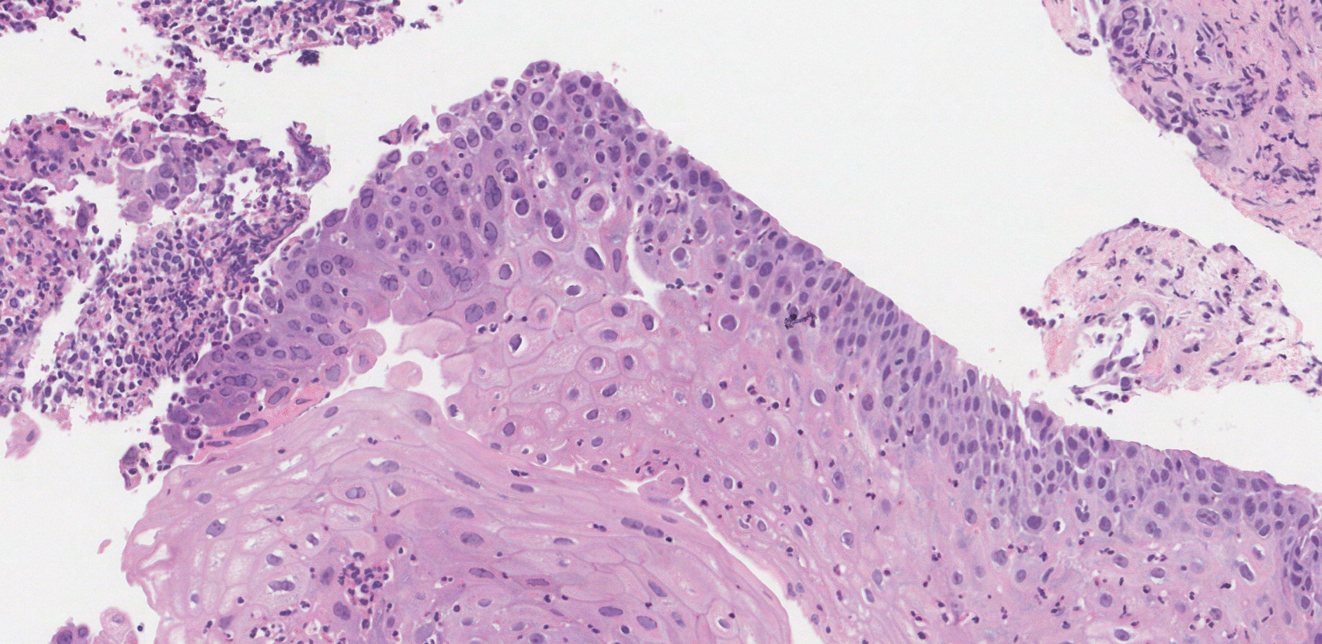
Microscopic features
Under the microscope, herpes esophagitis shows damage to the squamous cells that line the inside of the esophagus. This damage typically appears as ulcers (open sores). At the edge of these ulcers, pathologists may see infected squamous cells that look abnormal. Specifically, infected cells often have enlarged nuclei (the cell’s control center), and the chromatin (genetic material) inside the nuclei may be pushed to the edge. These cells might also contain multiple nuclei (called multinucleated cells) or characteristic intranuclear inclusions (small blue or purple spots inside the nucleus). Collectively, these changes are described as viral cytopathic effects. The area around these infected cells usually contains inflammatory cells, such as lymphocytes and neutrophils, which help fight infection.
What other tests may be performed to confirm the diagnosis?
Your pathologist may also use a special test called immunohistochemistry (IHC). This test helps confirm the presence of the herpes simplex virus by highlighting infected cells when viewed under the microscope.
We are proud to partner with:
![]()