

by Catherine Forse MD FRCPC
December 13, 2023
High grade squamous intraepithelial lesion (HSIL) is a precancerous disease caused by human papillomavirus (HPV). It is composed of abnormal squamous cells that have been infected and transformed by the virus. Squamous cells are found in a thin layer of tissue called the epithelium which covers the inside surface of the anus and anal canal. There are many types of HPV but most cases of HSIL are caused by the high-risk types 6, 11, 16, 18, and 51. Another name for HSIL of the anal canal is anal intraepithelial neoplasia (AIN).
HSIL is not cancer although patients with HSIL are at increased risk for developing a type of anal cancer called squamous cell carcinoma. For this reason, most patients with HSIL are offered treatment to remove the area of abnormal tissue. Low-grade squamous intraepithelial lesion (LSIL) is a related condition that is also caused by HPV. However, compared to HSIL, the risk of developing cancer from LSIL is much lower.
Can the cells in high grade squamous spread to other parts of the body?
No. Because HSIL is a non-invasive condition, the abnormal squamous cells are unable to spread to tissues outside of the anal canal or to other parts of the body such as lymph nodes.
How is this diagnosis made?
If your doctor is concerned that you may have an abnormality of your anal canal, they will perform a procedure called anoscopy. During anoscopy, your doctor will insert an instrument called an anoscope into your anal canal. An anoscope has a light source that allows your doctor to see the entire inner surface of the anal canal. If there is an area of abnormal tissue, your doctor may take a small sample of tissue called a biopsy. In some hospitals, doctors may insert a special swab into the anal canal instead of performing a biopsy. The swab collects tissue cells from the surface of the anal canal. The specimen that is collected is called a cytology specimen. After tissue samples from the anal canal have been collected, they are examined under a microscope by a pathologist.
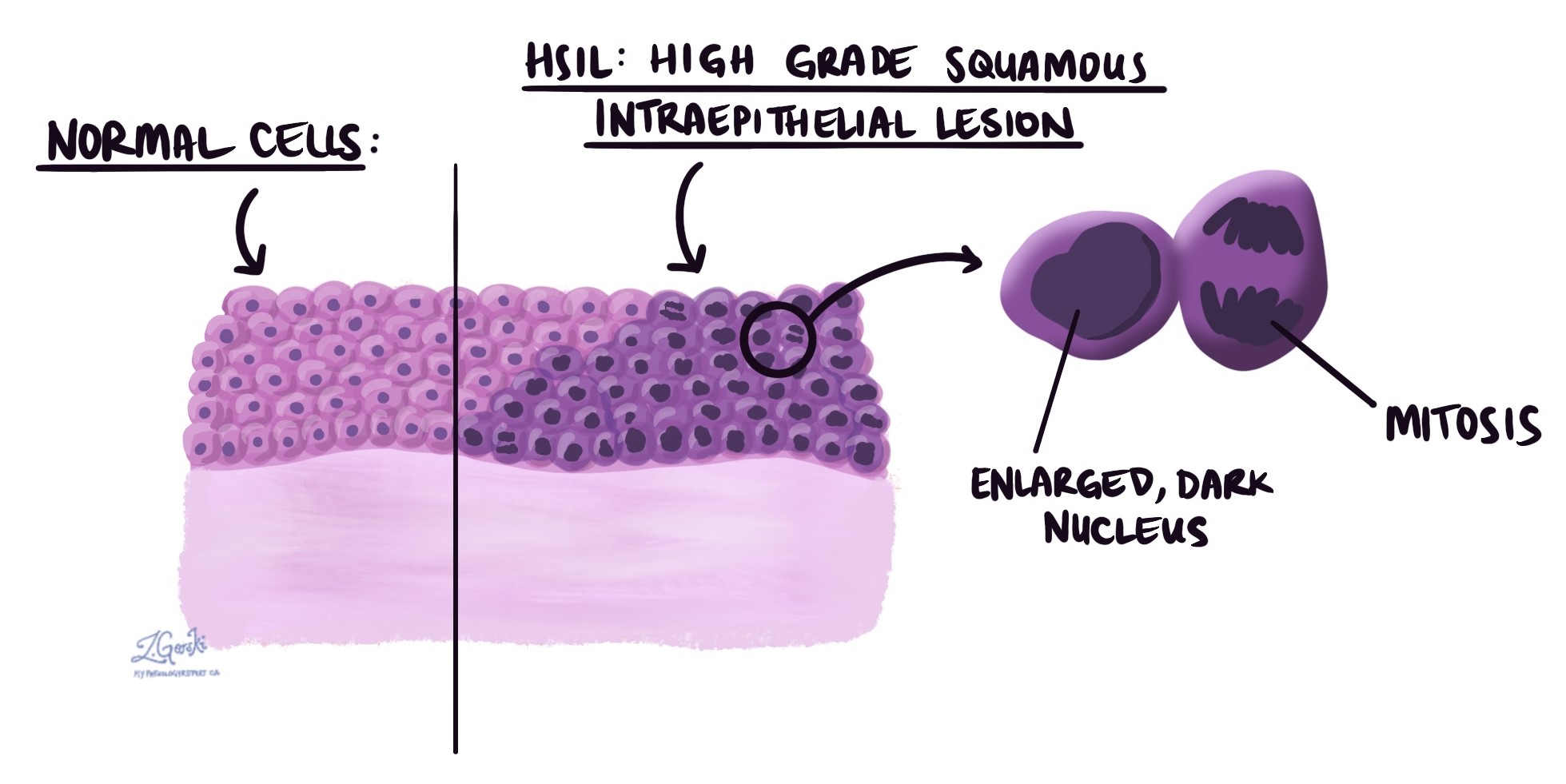
What does high grade squamous intraepithelial lesion look like under the microscope?
Under microscope examination, the abnormal squamous cells are confined to the epithelium on the surface of the tissue. The abnormal squamous cells are typically larger and darker than the surrounding normal squamous cells. Cells undergoing a process called mitosis (dividing to create new cells) are also usually seen. Pathologists often confirm the diagnosis by performing a test called immunohistochemistry (IHC) for a protein called p16. The abnormal cells in HSIL will be positive for p16 whereas other conditions that can look like HSIL will be negative.
What happens after high grade squamous intraepithelial lesion is diagnosed?
All patients with HSIL should be followed closely or offered treatment to remove the disease.
There are several treatment options available for HSIL:
- Local ablative therapy – Radiofrequency ablation or electrocautery can be used to remove the atypical tissue from the anal canal.
- Topical therapy – A medication (e.g. trichloroacetic acid, imiquimod, 5-fluorouracil) can be applied to specific lesions or to the entire anal canal.
- Surgery – Removal of the abnormal tissue through surgery may be recommended by your doctor if there is concern that there may also be squamous cell carcinoma.
Please talk to your doctor about which of the options is best suited to you.
About this article
This article was written by doctors to help you read and understand your pathology report. Contact us if you have questions about this article or your pathology report. For a complete introduction to your pathology report, read this article.
Other helpful resources
Canadian Cancer Society
We are proud to partner with:
![]()