

by Pavandeep Gill, MD FRCPC and Allison Osmond, MD FRCPC
June 8, 2023
What is seborrheic keratosis?
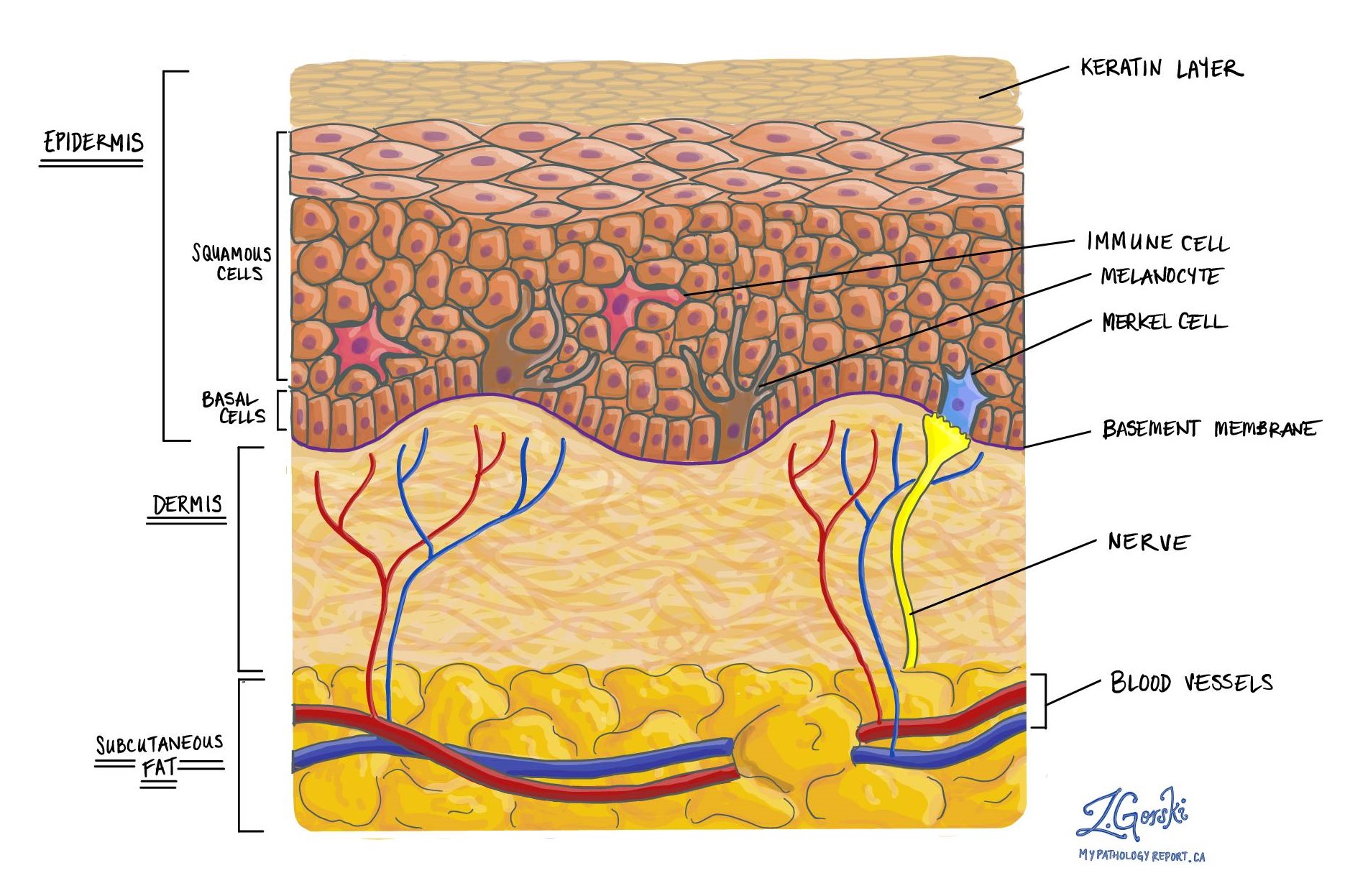
Seborrheic keratosis (SK) is a non-cancerous growth that starts from the squamous cells in the epidermis on the surface of the skin. Seborrheic keratosis is a very common condition and is seen more often as people age.
What does seborrheic keratosis look like?
Without a microscope, seborrheic keratosis may look like a raised or bumpy area of skin with a light tan or black colour. It is often described as looking as if it was “stuck on” to skin. The size of the involved skin can range from a few millimeters to a couple of centimeters and many patients have more than one. It may look similar to a wart or liver spot.
How do pathologists make this diagnosis?
The diagnosis is usually made after a small area of skin is removed in a procedure called an excisional biopsy or an excision. The tissue is then sent to a pathologist who examines it under a microscope.
What does seborrheic keratosis look like under the microscope?
When examined under the microscope, seborrheic keratosis is made up of small, round, and flattened squamous cells. Small spaces called cysts filled with keratin are also seen in the epidermis. Pathologists call these spaces “keratin pearls” because they are filled with rings of keratin. Your pathologist will examine the tissue sample carefully to make sure there is no sign of cancer or pre-cancerous disease.

Types of seborrheic keratosis
Pathologists divide seborrheic keratosis into types based on how the cells look under the microscope. Each type is called a variant.
The most common variants of seborrheic keratosis are:
- Acanthotic – When examined under the microscope, the epidermis in this variant is much thicker than in a regular seborrheic keratosis.,
- Papillomatous – In this variant, the cells at the surface grow in tall projections that pathologists describe as papillary.
- Adenoid/reticulated – Thin groups of squamous cells are seen extending into the deeper layers of the skin.
- Clonal – All of the squamous cells in this type have a very similar look as if they were all ‘cloned’ from a single cell.
- Inverted follicular keratosis – Instead of growing up from the surface, the abnormal cells in this type grow down towards the dermis.
- Inflammatory – In addition to the abnormal squamous cells, lots of immune cells are all seen in this type.
- Lichenoid – Many immune cells called lymphocytes are seen at the bottom of the epidermis, below the abnormal squamous cells.
- Desmoplastic – This type can look very similar to a type of cancer called squamous cell carcinoma. Although they look similar, desmoplastic seborrheic keratosis is still non-cancerous.
We are proud to partner with:
![]()