

by Jason Wasserman MD PhD FRCPC
July 1, 2026
Invasive breast carcinoma is the most common type of breast cancer. It begins in the cells lining the ducts and lobules of the breast and then spreads through the wall of a duct or lobule into the surrounding breast tissue. The word “invasive” means the cancer cells have broken out of the duct or lobule where they started and can now potentially spread to lymph nodes or other parts of the body.
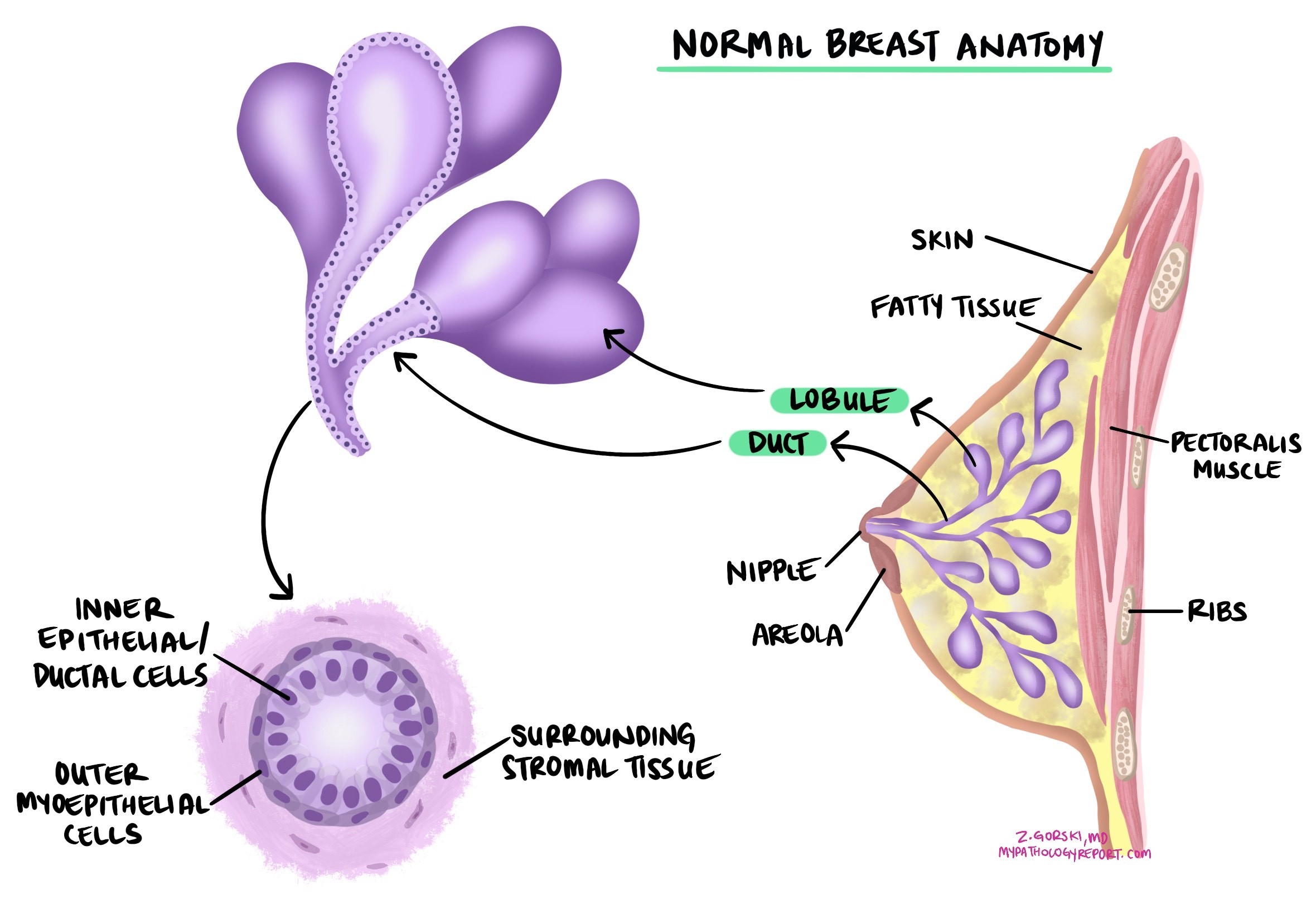
When a pathologist examines the tumor and it does not fit the pattern of a recognized special subtype, the cancer is called invasive breast carcinoma of no special type (NST). The older name for this cancer, still widely used in reports, is invasive ductal carcinoma. Pathologists make this diagnosis by excluding the special subtypes, which include invasive lobular carcinoma, tubular carcinoma, cribriform carcinoma, mucinous carcinoma, invasive micropapillary carcinoma, carcinoma with apocrine differentiation, and metaplastic carcinoma. Because “no special type” is a diagnosis of exclusion, it is the largest single group of breast cancers.
This article will help you understand the findings in your pathology report for invasive breast carcinoma, what each term means, and why it matters for your care.
What causes invasive breast carcinoma?
The exact cause of invasive breast carcinoma is not fully understood, but several factors are known to increase risk. These include hormonal, reproductive, lifestyle, and genetic influences.
Lifetime exposure to the hormone estrogen is one of the most important factors, which is why early menarche, late menopause, fewer pregnancies, older age at first childbirth, and shorter breastfeeding duration are all associated with higher risk. Hormone replacement therapy after menopause and alcohol consumption also raise risk, particularly for hormone receptor-positive cancers. Higher body weight after menopause, limited physical activity, higher breast density, and prior radiation therapy to the chest are additional contributors.
Genetics also plays a role. Inherited changes in genes such as BRCA1 and BRCA2 substantially increase the risk of developing breast cancer, and they tend to be linked to particular tumor types. BRCA1-related cancers are more often triple-negative (see the biomarker section below), while BRCA2-related cancers are more often hormone receptor-positive. Most breast cancers, however, occur in people with no identifiable inherited cause.
What are the symptoms of invasive breast carcinoma?
Many invasive breast carcinomas are found on a screening mammogram before they cause any symptoms. When symptoms do occur, the most common ones include:
- A new lump or mass in the breast, often firm and irregular, although some are soft or round.
- A change in the size, shape, or appearance of the breast.
- Skin changes such as dimpling, puckering, thickening, or redness.
- Nipple changes, including inversion (turning inward), discharge (especially if bloody), or scaling.
- Swelling of part or all of the breast, even when no distinct lump is felt.
- A lump or swelling in the lymph nodes under the arm or near the collarbone.
- Persistent pain in one area, although breast pain is more often caused by benign conditions.
How is the diagnosis made?
The diagnosis of invasive breast carcinoma is usually made after a small sample of the tumor is removed in a procedure called a core needle biopsy, most often performed because of an abnormal finding on a mammogram or a lump felt on examination. The tissue is sent to a pathologist, who examines it under the microscope.
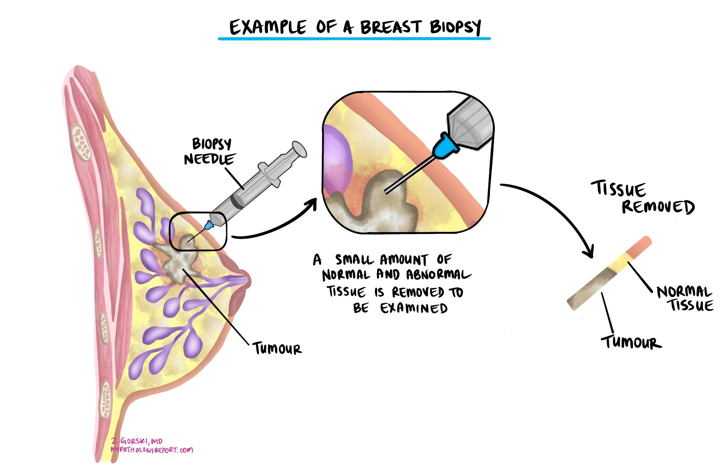
Under the microscope, the pathologist confirms invasive breast carcinoma by identifying cancer cells that have broken through the wall of the duct or lobule and are growing into the surrounding breast tissue. Non-invasive cancer (ductal carcinoma in situ) is often present alongside the invasive cancer, and the pathologist records both. When the tumor does not show the features of a recognized special subtype, it is classified as no special type.

The same tissue sample is also used to test the tumor for the hormone receptors estrogen receptor and progesterone receptor and for the protein HER2, which are described in the biomarker section below. After the biopsy confirms the diagnosis, surgery is usually performed to remove the tumor completely, and imaging such as ultrasound, MRI of the breast, or a mammogram is used to assess the size and extent of the tumor. If the cancer is large or has features that suggest a higher chance of spread, doctors may use additional imaging (such as CT or a bone scan) to check other parts of the body.
Histologic grade
Invasive breast carcinoma is given a histologic grade using the Nottingham grading system, which describes how closely the cancer cells resemble normal breast tissue and how quickly they appear to be growing. The grade helps predict how the cancer is likely to behave and is one factor used to guide treatment decisions. The pathologist scores three features, each from 1 to 3:
- Tubule formation — How much of the tumor is made up of normal-looking gland-like structures called tubules. More tubule formation is scored lower.
- Nuclear pleomorphism — How abnormal the cancer cell nucleus (the part of the cell that holds the DNA) looks compared with a normal cell, and how much the cells vary from one another. More abnormal nuclei are scored higher.
- Mitotic rate — How many cells are actively dividing. A higher number of mitotic figures is scored higher.

The three scores are added together to give a final grade:
- Grade 1 (low grade) — Total score 3 to 5. These tumors tend to grow slowly and are less likely to spread to lymph nodes or other parts of the body.
- Grade 2 (intermediate grade) — Total score 6 or 7. These tumors grow at a moderate pace and carry an intermediate risk of spread.
- Grade 3 (high grade) — Total score 8 or 9. These tumors tend to grow and spread more quickly.
Tumor size
The size of an invasive breast carcinoma is measured in centimeters and is important for two reasons. It is used to determine the pathologic tumor stage (pT, described in the staging section below), and larger tumors are more likely to spread to lymph nodes and other parts of the body. The full and final size can only be measured after the entire tumor has been removed at surgery, so a tumor size may not appear on your report after a biopsy. When more than one separate tumor is present, the report usually describes the largest one.
Lymphovascular invasion
In invasive breast carcinoma, lymphovascular invasion means that cancer cells are found inside the small lymphatic channels or blood vessels near the tumor. These vessels can serve as pathways that allow cancer cells to travel to lymph nodes or distant parts of the body. Lymphovascular invasion can be identified only when the pathologist sees cancer cells within a vessel under the microscope.
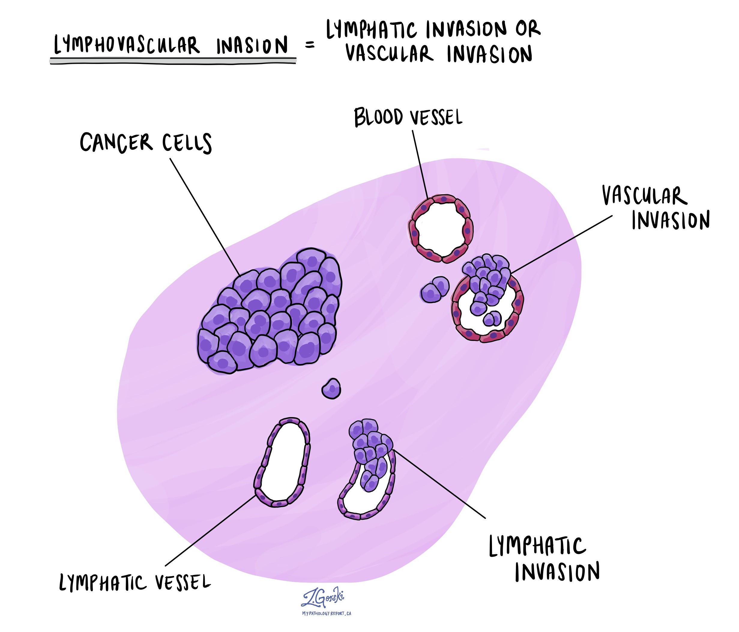
Lymphovascular invasion is an important prognostic finding in invasive breast carcinoma. It is associated with a higher risk that the cancer has spread to lymph nodes and a higher risk of recurrence, and it may influence decisions about additional treatment such as chemotherapy or radiation. Your report will usually describe lymphovascular invasion as “present” or “not identified.”
Margins
A margin is the edge of the tissue that the surgeon cuts to remove the tumor from the breast. In invasive breast carcinoma, margins are examined to determine whether the entire tumor was removed or whether some cancer may have been left behind. Margins are usually only reported after a surgical procedure to remove the whole tumor (a lumpectomy or mastectomy), not after a biopsy that removes only part of it.
- Negative margin — No cancer cells are seen at the cut edge of the tissue. This suggests the tumor was completely removed. Many reports also state how far the closest cancer cells are from the edge.
- Close margin — Cancer cells are near the cut edge but do not reach it. Depending on the distance, the team may discuss further surgery or radiation.
- Positive margin — Cancer cells are present at the cut edge, which means some tumor may remain in the breast. A positive margin may lead to another surgery to remove the remaining tumor or additional radiation to that area.

Lymph nodes
Lymph nodes are small, bean-shaped immune organs that filter fluid from the tissues. In invasive breast carcinoma, cancer cells often travel first to the lymph nodes under the arm (the axillary lymph nodes) before reaching other parts of the body, so examining these nodes is one of the most important ways to learn whether the cancer has started to spread. This information helps stage the cancer, estimate prognosis, and guide treatment decisions.
To check the nodes, surgeons often first remove one or a few sentinel lymph nodes, the first nodes cancer cells are most likely to reach. If these nodes contain cancer, surgeons may remove more nodes. Your report will typically include:
- The total number of lymph nodes examined and how many contain cancer.
- The size of the largest cancer deposit within a node.
- Whether extranodal extension is present, meaning cancer cells have broken through the outer capsule of a lymph node into the surrounding tissue.
The size of the largest deposit is described using three terms:
- Isolated tumor cells — A group of cancer cells 0.2 mm or smaller. Nodes with only isolated tumor cells are not counted as positive for the nodal stage and are recorded as pN0(i+).
- Micrometastasis — A deposit larger than 0.2 mm but not larger than 2 mm. When only micrometastases are found, the nodal stage is pN1mi.
- Macrometastasis — A deposit larger than 2 mm. Macrometastases are associated with a higher risk of recurrence and may lead to additional treatment.
Biomarker and molecular testing
Biomarker testing is an essential part of every invasive breast carcinoma workup, because the results define the type of breast cancer and determine which treatments are likely to work. Every invasive breast carcinoma is tested for three core markers: the estrogen receptor (ER), the progesterone receptor (PR), and HER2. Together, these three results place the cancer into a subtype that shapes the entire treatment plan. Additional molecular tests may be performed depending on the situation.
Estrogen receptor (ER) and progesterone receptor (PR)
Estrogen receptor (ER) and progesterone receptor (PR) are proteins found inside the nucleus of some breast cancer cells. When estrogen and progesterone attach to these receptors, they can signal cancer cells to grow. A cancer with ER or PR is called hormone receptor-positive.
Hormone receptor status is tested by immunohistochemistry (IHC) and reported as the percentage of cancer cells that show the receptor, along with the staining strength (intensity). A cancer is considered hormone receptor-positive when ER or PR is present in 1% or more of the cancer cells. Results between 1% and 10% are reported as ER-low positive, a category that behaves differently from strongly positive cancers and is managed accordingly. Some reports also give a combined score such as the Allred score or H-score.
Hormone receptor status indicates whether hormone (endocrine) therapy is likely to be effective. Hormone receptor-positive cancers can be treated with drugs that block estrogen or lower its levels, such as tamoxifen or aromatase inhibitors (anastrozole, letrozole, exemestane). Hormone receptor-positive cancers also tend to grow more slowly and generally have a more favorable outlook than hormone receptor-negative cancers.
HER2
HER2 (human epidermal growth factor receptor 2) is a protein on the surface of breast cells that helps control growth and division. In some invasive breast carcinomas, the HER2 gene is amplified (too many copies are made), and the cells produce far too much HER2 protein, which drives the cancer to grow. HER2 status determines whether HER2-targeted drugs are likely to be effective, so it is tested in every invasive breast carcinoma. You can read more in our article on HER2 in breast cancer.
HER2 testing follows a two-step process. Immunohistochemistry (IHC) is performed first and is scored from 0 to 3+. When the IHC result is 2+ (equivocal), fluorescence in situ hybridization (FISH) quantifies HER2 gene copy number and resolves the result. The result is reported in one of these categories:
- HER2-positive — IHC 3+, or IHC 2+ with FISH showing gene amplification. These cancers respond to HER2-targeted therapies such as trastuzumab, pertuzumab, and trastuzumab-deruxtecan.
- HER2-low — IHC 1+, or IHC 2+ with FISH not amplified. These cancers carry a small amount of HER2 protein. They are not HER2-positive in the traditional sense, but they may respond to the antibody-drug conjugate trastuzumab-deruxtecan in the metastatic setting. Because of this, pathologists now report the difference between IHC 0 and IHC 1+.
- HER2-ultralow — IHC 0 with faint, incomplete membrane staining in some cells. This newer category may also be eligible for trastuzumab-deruxtecan in hormone receptor-positive metastatic disease.
- HER2-negative — IHC 0 with no staining. HER2-targeted therapies are not effective.
Molecular subtype
The combination of ER, PR, and HER2 places invasive breast carcinoma into a molecular subtype, which is one of the strongest factors in treatment planning:
- Hormone receptor-positive, HER2-negative — The most common subtype. Usually treated with hormone-blocking therapy, often with surgery and radiation, and sometimes chemotherapy depending on the risk of recurrence.
- HER2-positive — Treated with HER2-targeted therapy combined with chemotherapy, plus hormone therapy if the cancer is also hormone receptor-positive.
- Triple-negative — ER-negative, PR-negative, and HER2-negative. Does not respond to hormone therapy or HER2-targeted drugs and is treated primarily with chemotherapy, sometimes combined with immunotherapy.
Ki-67
The Ki-67 labeling index is the percentage of cancer cells that are actively dividing. A higher Ki-67 index indicates a faster-growing tumor. In hormone receptor-positive, HER2-negative breast cancer, Ki-67 is one of several factors that may influence whether chemotherapy is considered, although genomic tests (described below) now provide more precise information for many patients.
Genomic (gene expression) tests
For many hormone receptor-positive, HER2-negative cancers, a genomic test that measures the activity of a panel of genes helps estimate recurrence risk and whether chemotherapy is likely to add benefit beyond hormone therapy. These tests are usually ordered by the treating oncologist, and the results often come separately from the pathology report:
- Oncotype DX (21-gene recurrence score) — Reports a score that estimates recurrence risk and helps decide whether chemotherapy should be added in early hormone receptor-positive, HER2-negative breast cancer.
- MammaPrint (70-gene signature) — Classifies the cancer as low risk or high risk of recurrence.
- Prosigna (PAM50) — Reports an intrinsic subtype and a risk-of-recurrence score.
Additional biomarkers in advanced or metastatic disease
When invasive breast carcinoma is advanced or has spread, comprehensive molecular testing (often using next-generation sequencing) may look for changes that open the door to specific targeted or immune therapies:
- PD-L1 — In triple-negative breast cancer, PD-L1 testing by immunohistochemistry (reported as a Combined Positive Score, with a CPS of 10 or higher generally required) helps determine eligibility for the immunotherapy drug pembrolizumab combined with chemotherapy. You can read more in our overview of PD-L1 testing in cancer.
- PIK3CA, AKT1, and PTEN — Changes in these genes affect a growth pathway called PI3K/AKT. In hormone receptor-positive, HER2-negative advanced disease, a PIK3CA change may indicate benefit from a PI3K inhibitor (alpelisib or inavolisib), and a PIK3CA, AKT1, or PTEN change may indicate benefit from the AKT inhibitor capivasertib.
- ESR1 — A change in the ESR1 gene is a common reason that hormone-receptor-positive cancers stop responding to certain endocrine therapies. It may indicate a switch to an oral drug called elacestrant.
- BRCA1 and BRCA2 — An inherited (germline) change in BRCA1 or BRCA2 in a HER2-negative cancer may indicate benefit from a PARP inhibitor such as olaparib or talazoparib. A BRCA change may also point to a hereditary cancer syndrome that affects family members, so a positive result is usually followed by referral to a genetic counselor.
- Tumor-agnostic markers — Rarely, breast cancers carry markers that qualify for treatments approved across cancer types regardless of where the cancer started, including mismatch repair deficiency (dMMR) or high microsatellite instability (MSI-high), a high tumor mutational burden, or an NTRK gene fusion.
For more information, visit our Biomarkers and Genetic Testing section.
Response to treatment given before surgery (residual cancer burden)
Some patients with invasive breast carcinoma receive chemotherapy, HER2-targeted therapy, or hormone therapy before surgery. This is called neoadjuvant therapy. When surgery is done afterward, the pathologist examines how much cancer remains, because the amount of remaining cancer is a strong predictor of long-term outcome. When no invasive cancer remains in the breast or lymph nodes, this is called a pathologic complete response and is associated with the best outcomes, especially in HER2-positive and triple-negative breast cancers.
When cancer remains, many centers report the residual cancer burden (RCB) index, which combines the size of the tumor bed, the percentage of it still containing cancer, the amount of non-invasive (in situ) cancer, and the number and size of lymph node deposits into a single score. The result is grouped into four categories:
- RCB-0 — No residual invasive cancer (pathologic complete response). Best outcomes.
- RCB-I — Minimal residual cancer.
- RCB-II — A moderate amount of residual cancer.
- RCB-III — Extensive residual cancer.
A higher RCB category is associated with a higher risk of recurrence and may lead the team to consider additional treatment after surgery.
Pathologic stage (pTNM)
Invasive breast carcinoma is staged using the TNM system from the American Joint Committee on Cancer (AJCC), 8th edition. TNM stands for Tumor (T, the size and extent of the tumor), Nodes (N, whether cancer has reached the lymph nodes), and Metastasis (M, whether cancer has spread to distant parts of the body). The letter “p” means the stage is based on what the pathologist measured in the removed tissue. Imaging, rather than the pathologist, determines distant metastasis (M). For breast cancer, the AJCC 8th edition also defines a prognostic stage that combines anatomic TNM with histologic grade and ER, PR, and HER2 results, so two cancers of the same size can have different stages depending on their biology.
Tumor stage (pT)
- pT1 — Tumor 2 cm (20 mm) or smaller. This is divided into pT1mi (1 mm or smaller), pT1a (more than 1 mm up to 5 mm), pT1b (more than 5 mm up to 10 mm), and pT1c (more than 10 mm up to 20 mm).
- pT2 — Tumor larger than 2 cm but not larger than 5 cm.
- pT3 — Tumor larger than 5 cm.
- pT4 — Tumor of any size that has grown into the chest wall or skin. pT4a is growth into the chest wall; pT4b is growth into the skin causing ulcers or swelling; pT4c is both; pT4d is inflammatory breast cancer, in which the skin is red and swollen.
Nodal stage (pN)
- pN0 — No cancer in the lymph nodes. When only isolated tumor cells are found, the stage is recorded as pN0(i+).
- pN1 — Cancer in 1 to 3 axillary lymph nodes, or micrometastases only (pN1mi), or spread to internal mammary nodes detected on sentinel node biopsy.
- pN2 — Cancer in 4 to 9 axillary lymph nodes, or in internal mammary nodes without axillary node involvement.
- pN3 — Cancer in 10 or more axillary lymph nodes, in nodes below or above the collarbone, or in a combination of internal mammary and axillary nodes.
What is the prognosis for a person diagnosed with invasive breast carcinoma?
The prognosis for invasive breast carcinoma depends on how far the cancer has spread and on the biology of the tumor. Overall, outcomes are favorable, particularly when the cancer is found early. The five-year relative survival for cancer that is still confined to the breast is over 95%, and it remains high (around 85%) when the cancer has spread only to nearby lymph nodes. Survival is lower when the cancer has spread to distant organs. Several features on the pathology report influence outlook:
- Stage — Smaller tumors confined to the breast with no lymph node involvement have the most favorable outlook. Lymph node or distant organ involvement is associated with lower survival.
- Grade — Higher-grade (grade 3) tumors tend to grow and spread more quickly than lower-grade tumors.
- Hormone receptor and HER2 status — Hormone receptor-positive cancers generally have a more favorable outlook and can be treated with hormone therapy. HER2-positive cancers respond well to HER2-targeted therapy. Triple-negative cancers are more difficult to treat and tend to have a less favorable outlook.
- Lymphovascular invasion — Cancer cells in blood or lymphatic vessels are associated with a higher risk of spread and recurrence.
- Margin status — Complete removal with negative margins is associated with a lower risk of the cancer returning in the breast.
- Genomic test results — For hormone receptor-positive, HER2-negative cancers, a genomic recurrence score helps refine the estimate of recurrence risk.
- Response to neoadjuvant therapy — When treatment is given before surgery, a pathologic complete response (RCB-0) predicts the best outcome, while a higher residual cancer burden predicts a higher risk of recurrence.
What happens after this diagnosis?
After a diagnosis of invasive breast carcinoma, a team usually coordinates care and may include a breast surgeon, a medical oncologist, a radiation oncologist, a pathologist, a radiologist, and, when inherited risk is a concern, a genetic counselor. The pathology findings guide which options the team considers, rather than dictating a single path.
- Surgery — Most patients have surgery to remove the tumor, either breast-conserving surgery (lumpectomy) or removal of the whole breast (mastectomy). The choice depends on tumor size and location, margin results, and patient preference. Doctors usually sample lymph nodes at the same time.
- Radiation therapy — Often considered after breast-conserving surgery, and sometimes after mastectomy, to lower the risk of the cancer returning in the breast or chest wall.
- Hormone (endocrine) therapy — Considered when the cancer is hormone receptor-positive, to block or lower estrogen.
- HER2-targeted therapy — Considered when the cancer is HER2-positive, usually combined with chemotherapy.
- Chemotherapy — Considered based on stage, grade, subtype, and genomic test results, and given before or after surgery.
- Immunotherapy — May be considered for triple-negative breast cancer, particularly when PD-L1 is positive.
After treatment, follow-up includes regular examinations and imaging to watch for recurrence, along with attention to bone health, menopausal symptoms, and, when relevant, fertility and genetic counseling.
Questions to ask your doctor
- What type and subtype of breast cancer do I have?
- What is the size and grade of my tumor?
- Are my hormone receptor (ER and PR) results positive or negative, and what percentage of cells were positive?
- Is my cancer HER2-positive, HER2-low, HER2-ultralow, or HER2-negative?
- What molecular subtype is my cancer, and what does it mean for treatment?
- Was lymphovascular invasion present?
- Were my surgical margins negative, close, or positive?
- How many lymph nodes were examined, and how many contained cancer?
- Was extranodal extension present in any lymph node?
- What is my pathologic stage (pT and pN)?
- Should I have a genomic test such as Oncotype DX, and how will the result affect my treatment?
- Do my results suggest I should have genetic testing for an inherited cause such as a BRCA change?
- If I had treatment before surgery, what was my residual cancer burden, and what does it mean?
- Which treatments are appropriate based on my pathology findings, and what are the next steps?
Related articles on MyPathologyReport.com
- Invasive Ductal Carcinoma of the Breast: Understanding Your Pathology Report
- Invasive Lobular Carcinoma of the Breast: Understanding Your Pathology Report
- Ductal Carcinoma In Situ (DCIS) of the Breast: Understanding Your Pathology Report
- Lobular Carcinoma In Situ (LCIS)
- Understanding Your Breast Biopsy Pathology Report
- HER2 in Breast Cancer
- PD-L1 Testing in Cancer
We are proud to partner with:


🔍 Search MyPathologyReport
Type what you see on your report — for example a diagnosis or test name