

Section Editor: Bibianna Purgina MD FRCPC
October 6, 2026
Chondrosarcoma is a type of bone cancer that forms from cartilage. Cartilage is the firm, smooth tissue that covers the ends of bones in joints and gives shape to the ear and nose. In chondrosarcoma, the cancer cells produce cartilage instead of bone.
Chondrosarcoma is one of the most common bone cancers in adults. It usually affects people between 40 and 70 years old and is uncommon in children. The most common locations are the pelvis, thigh bone, upper arm bone, shoulder blade, and ribs.
Chondrosarcomas range from slow-growing tumors that are almost always cured by surgery to fast-growing cancers that can spread. The most important finding in your pathology report is the grade, which describes where your tumor falls on that range.
This article explains how chondrosarcoma is diagnosed and what the grade, type, margins, and other findings in your pathology report mean.
What is the grade, and why does it matter?
The grade describes how abnormal the cancer cells look under the microscope. Conventional chondrosarcoma is given one of three grades, and the grade is the strongest predictor of how the tumor will behave:
- Grade 1. The cells look very close to normal cartilage cells and are only slightly crowded. These tumors grow slowly and very rarely spread to other parts of the body. They can come back in the same place if not completely removed.
- Grade 2. The tumor contains more cells, the cells look more abnormal, and some are dividing. These tumors are more likely to come back after surgery, and a minority spread, most often to the lungs.
- Grade 3. The tumor is crowded with very abnormal cells, many of them dividing, and areas of dead tumor tissue are common. These tumors grow faster and are the most likely to spread.
Grade 3 tumors are uncommon. Most chondrosarcomas are grade 1 or grade 2.
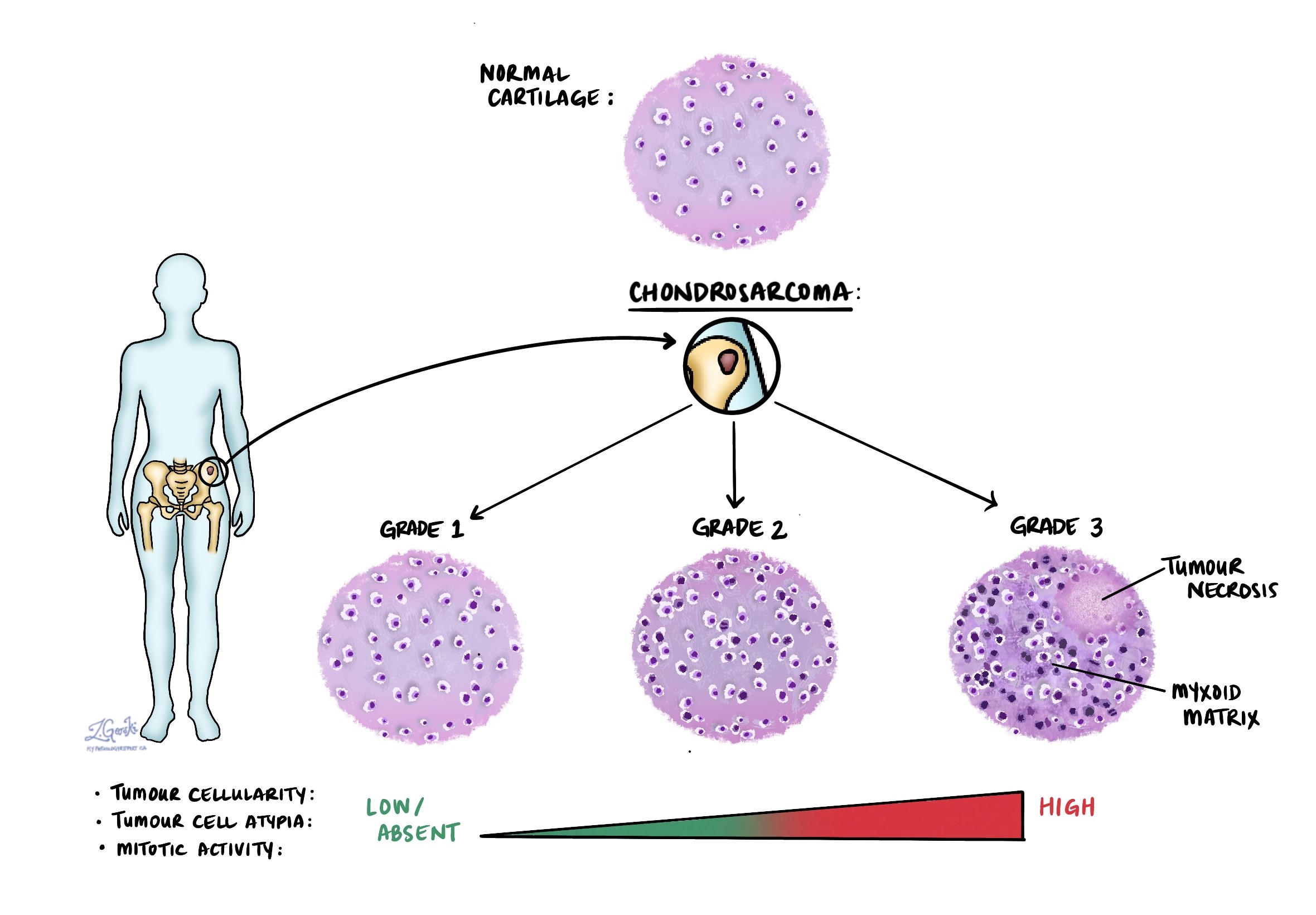
Grading a chondrosarcoma on a small biopsy can be difficult, because different parts of a tumor can look different. The grade is sometimes revised after the whole tumor has been removed and examined.
What is an atypical cartilaginous tumor?
When a low-grade cartilage tumor develops in the bones of the arms or legs, pathologists now call it an atypical cartilaginous tumor rather than grade 1 chondrosarcoma. The tumor looks the same under the microscope, but its location changes how it behaves.
In the arms and legs, these tumors rarely spread and can often be treated with a less extensive operation. The name grade 1 chondrosarcoma is kept for the same tumor when it develops in the pelvis, the shoulder blade, the spine, or the base of the skull. In those locations, complete removal is harder, and the tumor is more likely to come back.
Where in the bone does chondrosarcoma start?
Your report may describe the tumor as central or peripheral:
- Central chondrosarcoma. This type starts inside the bone, in the hollow center where the bone marrow is. It is the most common type. Some develop from a preexisting noncancerous cartilage tumor called an enchondroma.
- Peripheral chondrosarcoma. This type develops on the surface of a bone, usually from the cartilage cap of an osteochondroma, a common noncancerous bony outgrowth.
What are the special types of chondrosarcoma?
Most chondrosarcomas are the conventional type described above. A few less common types behave differently and are treated differently:
- Dedifferentiated chondrosarcoma. A low-grade chondrosarcoma in which part of the tumor has changed into a high-grade cancer that no longer makes cartilage. About 1 in 10 chondrosarcomas are this type. It grows quickly and spreads more often.
- Clear cell chondrosarcoma. A rare, slow-growing type that develops at the very end of a long bone, usually the top of the thigh bone or upper arm bone. It is low grade and has a good outlook when completely removed.
- Mesenchymal chondrosarcoma. A rare, fast-growing type that often affects teenagers and young adults. It contains small round cells and cartilage and is treated with chemotherapy and surgery.
- Periosteal chondrosarcoma. A rare type that grows on the outer surface of a bone. It is usually low grade.
What causes chondrosarcoma?
For most people, no cause is found. About half of central chondrosarcomas carry a change in a gene called IDH1 or IDH2. This change develops in the tumor cells during a person’s lifetime. It is not inherited and cannot be passed on to children.
A few conditions raise the risk of developing chondrosarcoma:
- Multiple enchondromas. People with many enchondromas (a condition called Ollier disease) or enchondromas and blood vessel growths (called Maffucci syndrome) have a higher risk. These conditions are caused by an IDH change that occurs early in development. They are not inherited.
- Multiple osteochondromas. This inherited condition causes many bony outgrowths and is caused by a change in the EXT1 or EXT2 gene. A small proportion of these outgrowths develop into peripheral chondrosarcoma. Each child of an affected parent has a 50% chance of inheriting it.
- Previous radiation therapy. Rarely, chondrosarcoma develops in a bone treated with radiation years earlier.
What are the symptoms of chondrosarcoma?
The most common symptom is a deep, aching pain in the affected bone. The pain often develops slowly, may be worse at night, and does not settle with rest. Some people notice a lump or swelling. A tumor near a joint can limit movement.
Low-grade tumors may cause very few symptoms and are sometimes found by chance on an X-ray or MRI done for another reason. New pain in a known cartilage tumor is a reason to have it checked.
How is the diagnosis made?
X-rays, CT, and MRI show a tumor that contains cartilage, often with small flecks of calcium. Imaging also shows whether the tumor is wearing away the bone or breaking through it, which helps separate a chondrosarcoma from a noncancerous enchondroma.
A biopsy confirms the diagnosis. The biopsy should be done at the center that will perform the final surgery, because the track left by the needle has to be removed along with the tumor later.
Separating a low-grade chondrosarcoma from an enchondroma is one of the hardest decisions in bone pathology, because the cells look very similar. Your pathologist relies as much on imaging as on the microscope, looking for signs that the tumor is growing into the surrounding bone.
Molecular testing
Testing for an IDH1 or IDH2 change may be done on the tumor. Finding one supports the diagnosis of a cartilage tumor and helps separate chondrosarcoma from chondroblastic osteosarcoma, a different bone cancer that also contains cartilage but is treated with chemotherapy.
An IDH change does not tell the pathologist whether a cartilage tumor is noncancerous or cancerous, because enchondromas carry the same change. IDH1 testing is also used to find people who may be eligible for clinical trials of targeted drugs, described below.
Tumor extension
Your report may describe whether the tumor has broken through the bone’s outer shell onto surrounding muscle or other soft tissue. This is called extraosseous extension.
The report may also note whether the tumor has grown into a nearby joint or an adjacent bone. In the spine and pelvis, which are made of several connected bones, spread from one bone to another raises the stage.
Margins
A margin is the edge of tissue cut by the surgeon. For chondrosarcoma, margins are especially important because surgery is the main treatment, and tumor left behind is the main reason the cancer comes back.
- Negative margin. No cancer cells are seen at the cut edge. The report may give the distance between the tumor and the closest margin.
- Positive margin. Cancer cells reach the cut edge, and some tumor may remain. This raises the chance the cancer will come back in the same place.
Your report will usually list several margins, including the cut ends of the bone, the surrounding soft tissue, and any large blood vessels or nerves that were removed.
Stage
Bone cancers are staged using the TNM system from the American Joint Committee on Cancer (AJCC), 8th edition. The tumor stage, called pT, depends on which bone is involved:
- Arms, legs, trunk, skull, and face. The stage depends mainly on size. A tumor 8 cm or smaller is pT1, and a tumor larger than 8 cm is pT2. Separate tumor deposits within the same bone are pT3.
- Spine and pelvis. These bones have their own staging rules, based on how many bone segments are involved and whether the tumor extends beyond the bone.
- Lymph nodes. Chondrosarcoma rarely spreads to lymph nodes, so surgeons usually do not remove them. When no nodes are examined, the report may say the nodal stage was not assigned.
When chondrosarcoma spreads, it usually travels through the bloodstream to the lungs.
What is the prognosis?
The outlook for chondrosarcoma depends most on the grade, the location, and whether the whole tumor can be removed. The figures below come from groups of people treated over many years and cannot predict what will happen to one person.
- Grade 1 and atypical cartilaginous tumor. Roughly 85% to 95% of people are alive five years after diagnosis, and almost all are cured.
- Grade 2. Roughly 70% to 85% of people are alive five years after diagnosis.
- Grade 3. The outlook is less favorable, and published figures vary widely with location and the completeness of surgery.
- Dedifferentiated chondrosarcoma. About 10% to 25% of people are alive five years after diagnosis.
Tumors in the pelvis and spine have a less favorable outlook than tumors in the limbs, mainly because they are harder to remove completely. Chondrosarcoma can come back many years after surgery, so follow-up continues for a long time.
What happens after the diagnosis?
A team at a center that regularly treats bone sarcomas manages chondrosarcoma.
- Surgery. Removing the tumor with a rim of normal tissue is the main treatment for most chondrosarcomas. Part of the bone may be replaced with an implant or graft.
- Atypical cartilaginous tumor in a limb. These may be treated by scraping out the tumor, a procedure called curettage, often followed by filling the space. Some small tumors are watched with imaging.
- Radiation therapy. Chondrosarcoma does not respond well to standard radiation. High-dose or proton radiation may be used when the tumor cannot be removed, particularly at the base of the skull.
- Chemotherapy. Standard chemotherapy works poorly for conventional chondrosarcoma and is not routinely used. It is used for mesenchymal chondrosarcoma and sometimes for dedifferentiated chondrosarcoma.
- Targeted treatment. For tumors with an IDH1 change that cannot be removed or have spread, a drug called ivosidenib is being studied in a large clinical trial. It is not yet approved for chondrosarcoma, and your oncologist can tell you whether you are eligible for a trial.
- Follow-up. Regular imaging of the original site and of the chest continues for many years.
Questions to ask your doctor
- What grade is my chondrosarcoma?
- Is my tumor called an atypical cartilaginous tumor or a chondrosarcoma, and why?
- Is it a conventional chondrosarcoma or one of the special types?
- Was the whole tumor removed, and were all the margins negative?
- Was the tumor tested for an IDH1 or IDH2 change?
- Has the cancer spread to my lungs or elsewhere?
- Do I need any treatment after surgery?
- How often will I need follow-up imaging, and for how many years?
Related articles on MyPathologyReport.com
We are proud to partner with:


🔍 Search MyPathologyReport
Type what you see on your report — for example a diagnosis or test name