

by Jason Wasserman MD PhD FRCPC
July 31, 2024
Background:
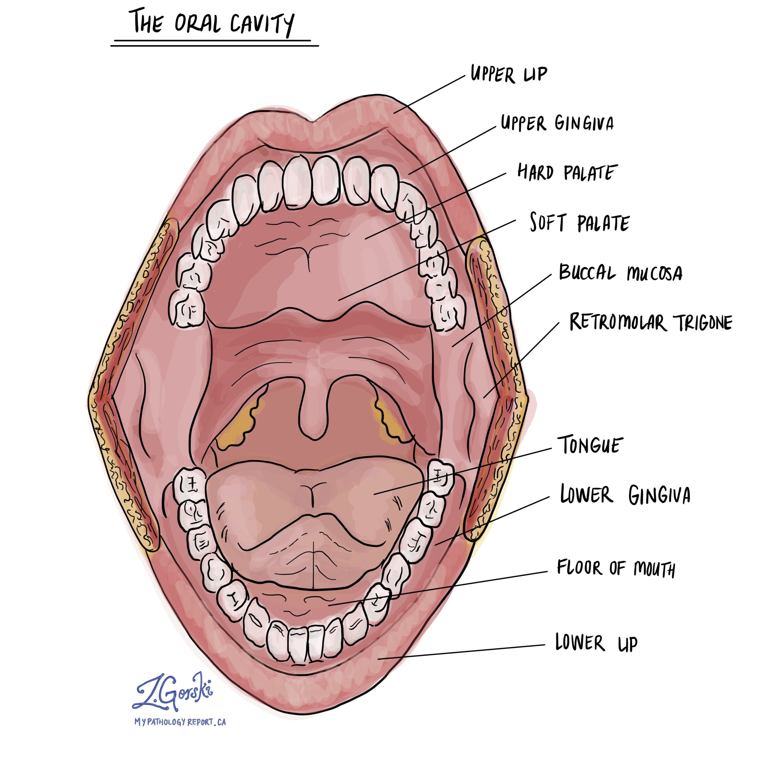
HPV associated dysplasia of the oral cavity is a precancerous condition in which the squamous cells that cover the inside of the oral cavity begin to show abnormal growth due to infection with the human papillomavirus (HPV). If not treated effectively, this condition can progress to a type of oral cavity cancer called squamous cell carcinoma. HPV associated dysplasia can affect any part of the oral cavity, although the tongue and floor of the mouth are most commonly affected.
What are the symptoms of HPV associated dysplasia of the oral cavity?
The condition may not present noticeable symptoms in the early stages, but possible signs can include pain, difficulty swallowing, unexplained lumps, or changes in the appearance of the oral mucosa. Lesions may appear slightly raised, white, or red in color.
What causes HPV associated dysplasia of the oral cavity?
HPV associated dysplasia is caused by infection with certain types of HPV, particularly the high-risk variants 16 and 18. These variants also cause squamous cell carcinoma of the oropharynx, cervix, and anal canal. HPV infects the squamous cells of the oral cavity and induces changes that can lead to dysplasia.
How is this diagnosis made?
The diagnosis of HPV associated dysplasia is typically made through a biopsy of the affected tissue. This sample is examined under a microscope to assess the presence and degree of dysplasia. Additionally, the presence of HPV RNA or DNA may be confirmed through molecular testing tests such as in situ hybridization (ISH) or polymerase chain reaction (PCR).
Microscopic features of HPV associated dysplasia
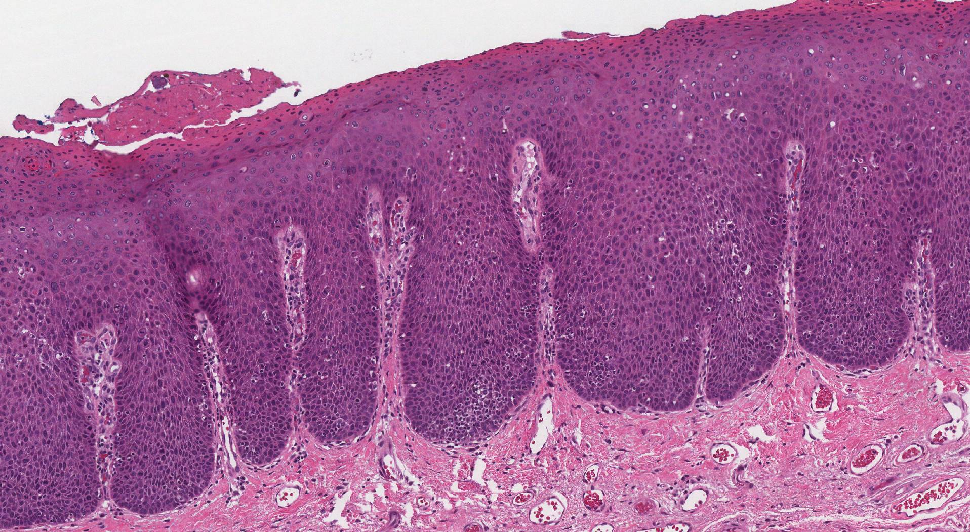
Microscopically, HPV associated dysplasia is characterized by abnormalities in the squamous cells, including pleomorphism (variation and cell size and shape), increased nuclear-to-cytoplasmic ratio, hyperchromatic nuclei, and possibly increased mitotic activity. The architectural organization of epithelium may also be disrupted. Pathologists often describe this type of dysplasia as nonkeratinizing because the abnormal squamous cells have not undergone a process called keratinization. This makes the abnormal squamous cells appear blue when examined under the microscope. In contrast, HPV independent squamous dysplasia looks pinker as the cells typically undergo keratinization.
Grading of HPV associated dysplasia
Unlike HPV-independent dysplasia, HPV associated dysplasia of the oral cavity is not graded because grade has not been shown to accurately correlate with the risk of developing cancer.
p16 testing in HPV associated dysplasia
p16 is a protein that is commonly overexpressed in cells affected by HPV. Pathologists test for p16 as a biomarker for HPV associated dysplasia and cancers because its presence strongly correlates with HPV infection. Overexpression of p16 in the cells of a biopsy is used to support the diagnosis of HPV-related pathology. The p16 test can be particularly useful as it helps differentiate HPV-associated lesions from other conditions that are not related to HPV.
What is the risk that HPV associated dysplasia will turn into cancer?
In the limited number of studies done to date, 10 to 15% of all patients diagnosed with HPV associated dysplasia went on to develop a type of oral cavity cancer called squamous cell carcinoma over time. Surgery to completely remove the area of dysplasia is associated with a lower risk and better overall prognosis.
About this article
Doctors wrote this article to help you read and understand your pathology report. If you have any questions, please contact us.
Learn more pathology
Atlas of Pathology
We are proud to partner with:
![]()