

by Catherine Forse MD FRCPC
March 30, 2026
Lymphocytic colitis is a non-cancerous condition in which an increased number of immune cells called lymphocytes accumulate in the colon’s lining, causing inflammation and damage. It belongs to a group of conditions called microscopic colitis, named because the changes that cause symptoms are invisible to the naked eye and can only be seen when tissue is examined under a microscope. The other main type of microscopic colitis is collagenous colitis, which shares many features with lymphocytic colitis.
Lymphocytic colitis is not cancer and does not increase the risk of developing cancer. It is a chronic condition for many people, but it responds well to treatment in most cases, and the outlook is generally good.
What are the symptoms?
The hallmark symptom of lymphocytic colitis is chronic watery diarrhea that can last for weeks, months, or years. The diarrhea develops because the accumulated lymphocytes damage the lining of the colon, impairing its ability to absorb water normally. Other symptoms may include abdominal cramping or pain, bloating, fatigue, and unintentional weight loss.
Symptoms often come and go. Some people experience prolonged flares followed by periods of improvement, while others have more persistent symptoms. The severity varies widely.
Who is affected?
Lymphocytic colitis is more common in middle-aged and older adults. Unlike collagenous colitis, which affects women significantly more often than men, lymphocytic colitis occurs at similar rates in both sexes. It is one of the more common causes of chronic watery diarrhea in adults over fifty, though it remains relatively uncommon overall.
What causes lymphocytic colitis?
The exact cause is not fully understood and likely involves a combination of factors. Several contributing causes have been identified:
- Medications. Certain drugs are strongly associated with lymphocytic colitis and may trigger or worsen it. The most commonly implicated include non-steroidal anti-inflammatory drugs (NSAIDs such as ibuprofen and naproxen), proton pump inhibitors (such as omeprazole and pantoprazole), selective serotonin reuptake inhibitors (SSRIs, a class of antidepressants), and olmesartan (a blood pressure medication). If a medication is identified as a likely trigger, stopping it often leads to significant improvement or complete resolution of symptoms.
- Autoimmune reaction. The immune system appears to play a central role. One leading theory is that lymphocytes mistakenly attack the cells lining the colon, triggering chronic inflammation.
- Changes in gut bacteria. Alterations in the normal bacterial environment of the colon may trigger an abnormal immune response in some people.
- Reaction to substances in the colon. Another theory suggests that the immune response may be triggered by material in stool passing through the colon, though the specific triggers have not yet been identified.
- Genetic factors. Some people may have a genetic predisposition that makes them more susceptible to developing microscopic colitis, though the specific genes involved are not well established.
In many cases, no single clear trigger is found, and the condition is managed based on symptoms rather than an identified cause.
How is the diagnosis made?
If your doctor suspects lymphocytic colitis based on your symptoms, they will recommend a colonoscopy — a procedure that uses a small flexible camera to look inside the colon. During the procedure, your doctor will take small tissue samples, called biopsies, from several parts of the colon. Taking biopsies from multiple locations is important because lymphocytic colitis can be patchy — it may affect one area of the colon. Still, not another, and a single biopsy from one site could miss the changes entirely.
In most cases, the colon looks completely normal to the camera during the colonoscopy. The diagnosis can only be confirmed when a pathologist examines the biopsies under a microscope and identifies the characteristic features described below.
What does the pathology report describe?
The pathologist looks for a specific pattern of changes in the colon lining. Unlike chronic active colitis associated with inflammatory bowel disease, lymphocytic colitis does not cause serious structural damage to the colon — it is a surface-level process.
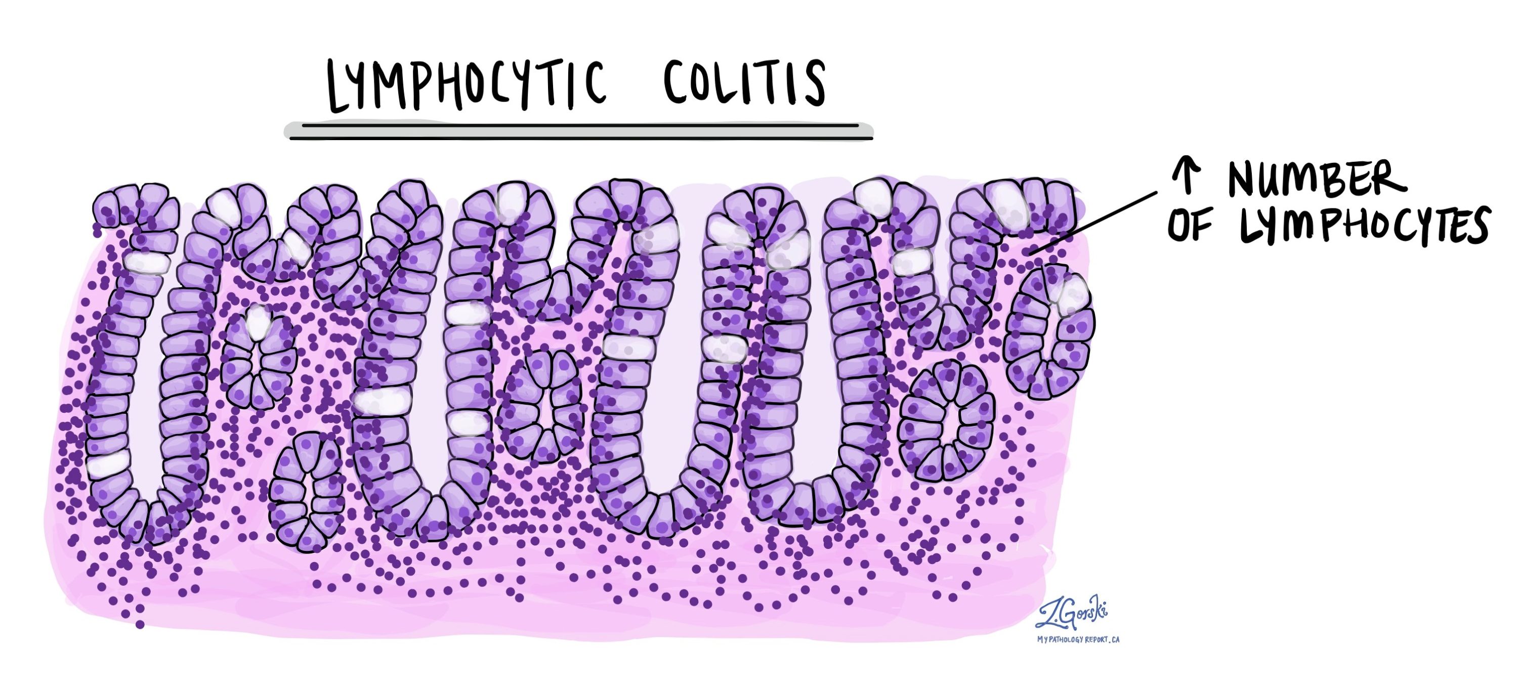
- Intraepithelial lymphocytosis. This is the defining feature of lymphocytic colitis. An abnormally large number of lymphocytes are found within the epithelium — the surface layer of cells that lines the colon — and in the lamina propria, the supportive tissue just beneath. Pathologists define intraepithelial lymphocytosis as more than 20 lymphocytes per 100 surface epithelial cells. In lymphocytic colitis, this count is typically much higher.
- Gland damage and atrophy. Over time, the accumulated lymphocytes damage the glands (also called crypts) that line the colon. The gland cells become smaller — a change pathologists call atrophy. Damaged glands produce less mucin, the substance that normally lubricates and protects the colon lining. This damage, combined with the surface inflammation, impairs the colon’s ability to absorb water, which is why watery diarrhea is the dominant symptom.
- Increased immune cells in the lamina propria. Beyond the lymphocytes in the epithelium, the lamina propria also contains an increased number of immune cells, indicating ongoing inflammation throughout the lining.
One important point: unlike ulcerative colitis and Crohn’s disease, lymphocytic colitis does not cause the structural changes — such as crypt distortion, ulcers, or abscesses — that are typical of inflammatory bowel disease. This distinction helps the pathologist confirm the diagnosis and is also reassuring: the colon has not sustained the kind of long-term architectural damage seen in IBD.
Lymphocytic colitis also differs from collagenous colitis in one key way: it does not show the thickened band of collagen beneath the surface that defines collagenous colitis. Both conditions share the intraepithelial lymphocytosis pattern, but the absence of a collagen band is what makes the diagnosis lymphocytic rather than collagenous colitis.
What happens next?
Lymphocytic colitis is a treatable condition, and most people experience significant improvement with appropriate management. The first step is to review any medications that may have triggered or worsened the condition. If an offending drug — particularly an NSAID, proton pump inhibitor, SSRI, or olmesartan — is identified, stopping it can lead to substantial improvement or even complete resolution of symptoms.
When medication adjustment alone is not enough, or when no medication trigger is identified, several treatments are effective:
- Budesonide. This is the most effective and most commonly used medication for lymphocytic colitis, as it is for collagenous colitis. It is a corticosteroid (anti-inflammatory drug) that works locally in the colon, with minimal systemic absorption, reducing the side effects associated with typical steroids. Most people respond well to a short course, though symptoms can return when it is stopped, and some patients require longer-term or intermittent treatment.
- Bismuth subsalicylate. This over-the-counter medication (the active ingredient in Pepto-Bismol) can reduce symptoms in milder cases and is sometimes used as an initial option.
- Diet and lifestyle adjustments. Some people find that reducing caffeine, alcohol, or dairy helps manage symptoms. These measures do not treat the underlying inflammation but can reduce the frequency of diarrhea.
- Other medications. In more persistent or severe cases, a gastroenterologist may consider additional options, including immunosuppressants or biologic therapies.
Many people with lymphocytic colitis experience periods of spontaneous improvement, and some go into remission without active treatment. Relapses are common, however, and ongoing follow-up with a gastroenterologist is important for managing the condition over time. Regular colonoscopies are not usually needed for surveillance purposes, since lymphocytic colitis does not increase the risk of colorectal cancer.
Questions to ask your doctor
- Could any of my current medications be contributing to this condition?
- What treatment do you recommend, and how long will I need to take it?
- What should I do if my symptoms return after finishing treatment?
- Are there dietary changes that might help manage my symptoms?
- Will I need follow-up colonoscopies, and if so, how often?
- Is there anything about my case that would suggest a different underlying diagnosis?
Related articles
We are proud to partner with:
![]()