By Jason Wasserman MD PhD FRCPC
November 26, 2024
This article is designed to help you understand your pathology report for peripheral T cell lymphoma. Each section explains an important aspect of the diagnosis and what it means for you.
What is peripheral T cell lymphoma?
Peripheral T cell lymphoma, NOS, is a type of cancer that starts in T cells, a part of the immune system. It is called “peripheral” because it typically involves mature T cells found in lymph nodes, blood, or tissues outside the thymus (where T cells are formed initially). The term NOS (“not otherwise specified”) means that it doesn’t fit into any other specific subtype of T cell lymphoma. This type of lymphoma is rare and tends to behave aggressively.
What are the symptoms of peripheral T cell lymphoma NOS?
The symptoms of peripheral T cell lymphoma NOS can vary depending on where it is located in the body.
Common symptoms include:
- Swollen lymph nodes, which may feel like lumps under the skin.
- Fever.
- Night sweats.
- Unexplained weight loss.
- Fatigue.
- Rash or skin nodules if the lymphoma involves the skin.
- Pain or discomfort if it affects organs or tissues.
What causes peripheral T cell lymphoma NOS?
The exact cause of peripheral T cell lymphoma NOS is not well understood. It occurs when T cells develop genetic changes that make them grow uncontrollably. These changes can be caused by unknown factors, but some cases have been linked to viral infections, such as the Epstein-Barr virus (EBV) or a weakened immune system.
How is this diagnosis made?
The diagnosis is made by examining a sample of tissue under the microscope. This tissue is usually taken from a swollen lymph node or affected organ during a biopsy. Additional tests, such as immunohistochemistry and flow cytometry, are performed to identify the specific types of cells and proteins in the tissue.
Microscopic features
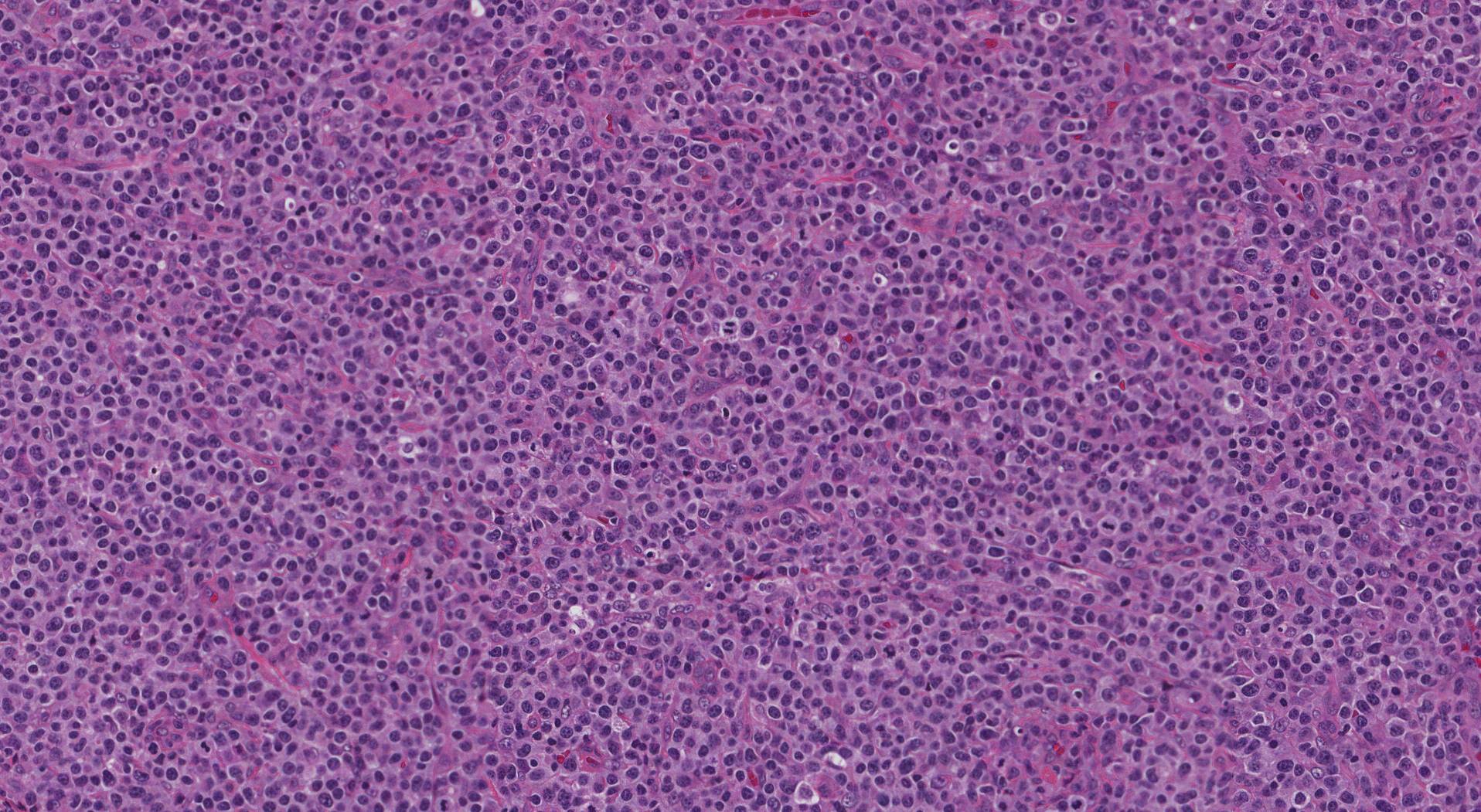
Pathologists use a microscope to examine the cells of the lymphoma. Peripheral T cell lymphoma, NOS, has a wide range of appearances, but some key features include:
- Clusters or sheets of abnormal T cells often replace the normal structure of the lymph node.
- These cells may vary in size and shape, often appearing medium to large with irregular shapes, darker-than-normal nuclei, and prominent nucleoli.
- In some cases, small to medium-sized T cells with clear cytoplasm dominate.
- The tissue often contains inflammatory cells, including small lymphocytes, plasma cells, eosinophils, and large B cells. The background inflammation can vary depending on the lymphoma’s molecular subtype.
- If the lymphoma involves the skin, it may form nodules in the deeper layers, sometimes affecting surrounding structures.

What other tests may be performed to confirm the diagnosis?
Immunohistochemistry and flow cytometry are specialized laboratory tests that help doctors understand more about lymphoma cells. These tests are essential for confirming the diagnosis and guiding treatment.
Immunohistochemistry
Immunohistochemistry is a test that uses special stains to highlight specific proteins on the surface of cells. These stains act like markers, helping pathologists identify the type of cells in a tissue sample. For example, this test can determine whether the lymphoma cells are T cells and whether they have markers typically seen in T cell lymphomas.
Flow cytometry
Flow cytometry is another type of test that analyzes individual cells. The cells are suspended in a liquid and passed through a laser beam, which measures the proteins on their surface. This test provides detailed information about the type and behavior of the lymphoma cells.
What do these tests show in peripheral T cell lymphoma, NOS?
In peripheral T cell lymphoma, not otherwise specified, the following patterns are often seen:
- The lymphoma cells typically have T cell markers, but some markers, such as CD5 and CD7, may be missing or reduced.
- Most cases show cells that are CD4-positive (a marker found on helper T cells) rather than CD8-positive (a marker found on cytotoxic T cells). However, rare cases may show both markers or neither.
- Some lymphoma cells produce proteins linked to cytotoxic T cells, such as TIA1 or granzyme B.
- In rare cases, the lymphoma cells may express proteins usually seen in B cells, such as CD20.
- A protein called CD30 may be present in some cases. This finding is important because it may influence treatment options.
These tests not only confirm that the lymphoma is a T cell lymphoma but also help identify specific features of the cancer. Knowing which markers are present can help doctors classify the lymphoma more precisely, predict how aggressive it might be, and choose the most effective treatment plan.
Prognosis
The prognosis for peripheral T cell lymphoma NOS depends on factors such as the stage of the disease, the patient’s overall health, and how well the lymphoma responds to treatment. In general, this type of lymphoma has an aggressive course, but treatment options, including chemotherapy, radiation, and stem cell transplant, can be effective for some patients. Targeted therapies, such as brentuximab vedotin for cases with CD30 expression, may also be an option in certain situations. Discussing your specific case and treatment options with your healthcare team is important.


