MyPathologyReport
September 21, 2023

Smooth muscle actin (SMA) is a protein that plays an important role in the structure and function of smooth muscle cells. Smooth muscle is one of the three types of muscle tissue found in the human body, with the other two being skeletal muscle and cardiac muscle.
SMA is a specialized protein that allows cells to contract (shorten) and relax (lengthen). As a result, cells that express SMA can be used to change the size of an organ or tissue or to open or close a channel such as a blood vessel.
SMA is expressed by smooth muscle cells throughout the body. These types of cells are normally found around blood vessels and in the walls of contractile organs such as the esophagus, stomach, small intestine, colon, and uterus. It is also expressed by other types of cells that behave like smooth muscle cells including myoepithelial cells, fibroblasts, and pericytes. These cells are normally found around glands, ducts, and soft tissue throughout the body.
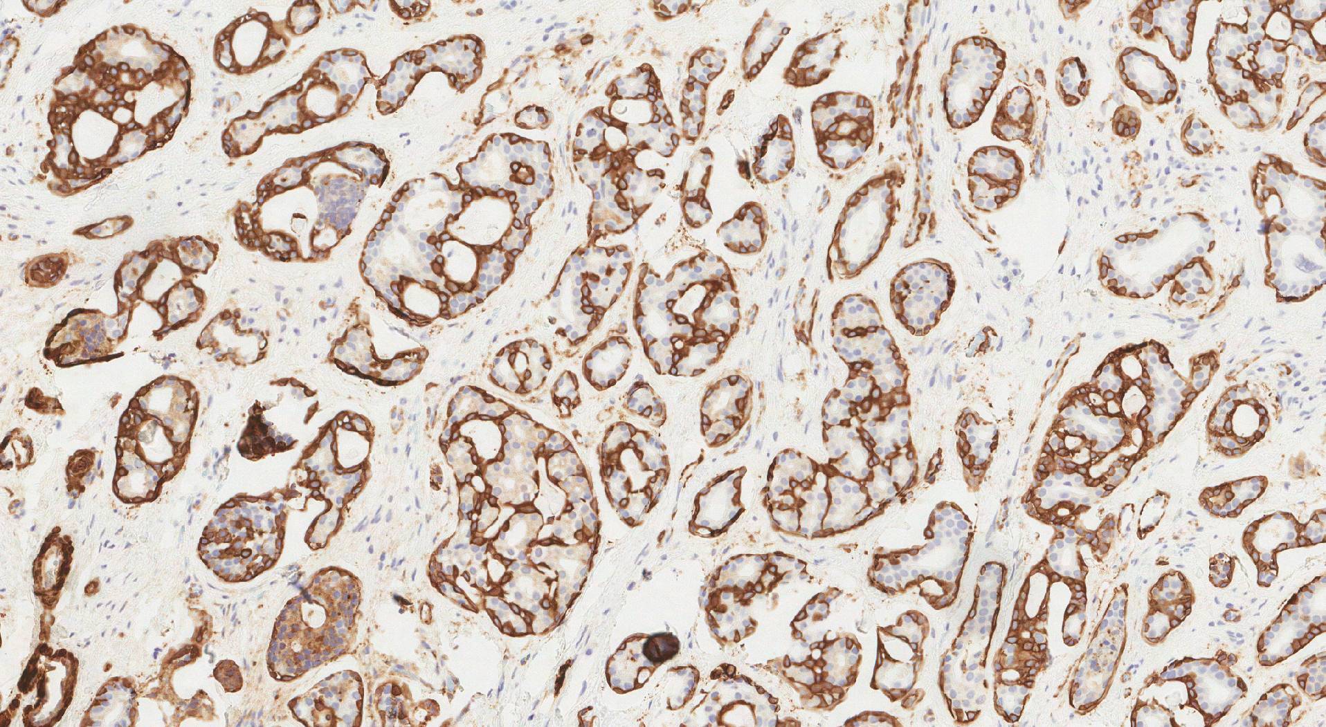
Pathologists can see SMA-producing cells by performing a test called immunohistochemistry (IHC). The test result is often reported as positive (the cells are making the protein) or negative (the cells are not making the protein).
Examples of tumours that are positive for SMA:
- Leiomyoma
- Leiomyosarcoma
- Myoepithelial cells
- Myoepithelioma
- Myoepithelial carcinoma
- Angioleiomyoma
- Pleomorphic adenoma
- Adenoid cystic carcinoma
- Epithelial-myoepithelial carcinoma
- Inflammatory myofibroblastic tumour (IMT)
- Fibromatosis
- Angiomyolipoma (PEComa)
- Gastrointestinal stromal tumour (GIST)
- Nodular fasciitis
- Synovial sarcoma
About this article
This article was written by doctors to help you read and understand your pathology report. Contact us if you have questions about this article or your pathology report. For a complete introduction to your pathology report, read this article.


